Multimedia Gallery
Formation of twins
Twins are rare and special, occurring in about 2% of all pregnancies. Of that number, 30% are identical twins. The other 70% are non-identical, or fraternal twins.
This animation will show you the differences between the development of a single baby, identical twins, and fraternal twins.
Starting with the single baby, let’s go back to the beginning, when fertilization occurs. Here you see that the egg cell is fertilized by a single sperm cell to form a zygote. Over the next few days, the fertilized egg cell divides over and over to form a structure composed of hundreds of cells called a blastocyst.
During the first week after fertilization, we can look inside the blastocyst and see the mass of cells that will form the embryo. The blastocyst will continue traveling toward the uterus where it will implant in the uterine lining, and grow into a single baby.
Now let’s watch the development of identical twins. Identical twins start out from a single fertilized egg cell, or zygote, which is why they’re also called monozygotic twins. Like the single baby we just saw, the egg cell is fertilized by a single sperm cell.
Unlike the single baby, this fertilized egg cell will split into two separate embryos, and grow into identical twins. This remarkable event takes place during the first week after fertilization, and can happen at several different times: at the two cell stage on day 2 at the early blastocyst stage on day 4 or in the late blastocyst stage on day 6.
The stage at which the egg cell splits determines how the twins will implant in the uterine lining, and whether or not they share an amnion, chorion, and placenta. Basically, the earlier the splitting occurs, the more independently the twins will develop in the uterus. So, a pair of identical twins that split during the two-cell stage will each develop its own amnion, chorion, and placenta.
Twins that split during the late blastocyst stage will share an amnion, chorion, and amniotic sac.
A common misconception about the conception of identical twins is that the trait for having them is passed on to future generations through the mother’s genes. But the truth is science doesn’t know the reason why identical twins occur. At this time, we can just say that they’re examples of a nine-month double miracle.
Now let’s take a look at the second type of twins. Non-identical, or fraternal, twins develop from two fertilized egg cells, or zygotes. Which is why they’re also called dizygotic twins. Unlike identical twins, however, fraternal twins are definitely influenced by the mother’s genes. Here’s why:
When the mother of fraternal twins ovulates, sometimes her ovaries release two egg cells for fertilization. Typically, only one egg cell is released during ovulation.
During conception, both of these egg cells become fertilized by two different sperm cells, which is why fraternal twins don’t look exactly alike. Sometimes they’re not even the same sex.
Here in the uterus, you can see that the twin embryos develop separately each having his or her own chorion, amnion, and placenta.
Formation of twins
Review Date: 8/12/2025
Reviewed By: LaQuita Martinez, MD, Department of Obstetrics and Gynecology, Emory Johns Creek Hospital, Alpharetta, GA. Also reviewed by David C. Dugdale, MD, Medical Director, Brenda Conaway, Editorial Director, and the A.D.A.M. Editorial team.
Animations
- Breast engorgement
- Cell division
- Cesarean section
- Conception - general
- Conception - pregnancy
- Conception of identical twins
- C-section
- Early labor
- Egg cell production
- Egg production
- Endometriosis
- Fetal ear development
- Formation of twins
- Human face formation
- Infant formulas
- Kids - How big is the baby?
- Kids - How does the baby co...
- Kids - Is it a girl or boy?
- Kids - Umbilical cord
- Kids - Where do babies come...
- Newborn jaundice
- NICU consultants and suppor...
- Ovulation
- Placenta delivery
- Placenta formation
- Preeclampsia
- Pregnancy
- Pregnancy care
- Sperm production
- Sperm release pathway
- Storing breast milk
- The role of amniotic fluid
- Twin-to-twin transfusion sy...
- Ultrasound
- Vaginal delivery
Illustrations
- 24-week fetus
- Abnormal discharge from the...
- Abnormal menstrual periods
- Absence of menstruation (am...
- Amniocentesis
- Amniocentesis
- Amniotic fluid
- Amniotic fluid
- Anatomy of a normal placenta
- Antibodies
- Baby burping position
- Bananas and nausea
- Blood cells
- Blood test
- Breast infection
- Breastfeeding
- Bulging fontanelles
- Candida - fluorescent stain
- Caput succedaneum
- Cesarean section
- Cesarean section
- Cesarean section
- Childbirth
- Chorionic villus sampling
- Congenital hip dislocation
- Congenital toxoplasmosis
- Crying - excessive (0 to 6 ...
- Delivery presentations
- Developmental milestones
- Early weeks of pregnancy
- Ectopic pregnancy
- Emergency Childbirth
- Emergency Childbirth
- Endocrine glands
- Endometriosis
- Endometritis
- Erythroblastosis fetalis - ...
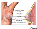
- Female breast
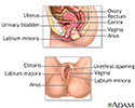
- Female reproductive anatomy
- Female reproductive anatomy
- Female reproductive anatomy...
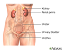
- Female urinary tract
- Fetal blood testing
- Fetal head molding
- Fetus at 10 weeks
- Fetus at 12 weeks
- Fetus at 16 weeks
- Fetus at 26 to 30 weeks
- Fetus at 3.5 weeks
- Fetus at 30 to 32 weeks
- Fetus at 7.5 weeks
- Fetus at 8.5 weeks
- First trimester of pregnancy
- Folic acid
- Folic acid benefits
- Folic acid source
- Follicle development
- Fontanelles
- Foreskin
- Gestational ages
- Gestational diabetes
- Gonadotropins
- Head circumference
- Heat rash
- Height/weight chart
- Hormonal effects in newborns
- Humidifiers and health
- Hysterectomy
- Infant blood sample
- Infant care following delivery
- Infant diaphragmatic hernia
- Infant heat rash
- Infant intestines
- Infant jaundice
- Infantile reflexes
- Influenza vaccines
- Intraductal papilloma
- Intrauterine transfusion
- Jaundiced infant
- Large fontanelles
- Large fontanelles (lateral view)
- Macrosomia
- Male reproductive anatomy
- Male reproductive anatomy
- Male urinary tract
- Mammary gland
- Meconium
- Morning sickness
- Moro reflex
- Newborn head molding
- Newborn test
- Normal female breast anatomy
- Normal uterine anatomy (cut...
- Ovarian cyst
- Ovarian hypofunction
- Overproductive ovaries
- Pelvic adhesions
- Pelvic laparoscopy
- Placenta
- Placenta
- Placenta
- Placenta previa
- Polyhydramnios
- Preeclampsia
- Pregnancy test
- Primary amenorrhea
- Primary infertility
- Secondary amenorrhea
- Secondary infection
- Side sectional view of fema...
- Single palmar crease
- Skull of a newborn
- Slit-lamp exam
- Sperm
- Stein-Leventhal syndrome
- Sunken fontanelles (superio...
- Tobacco health risks
- Transvaginal ultrasound
- Ultrasound in pregnancy
- Ultrasound, color - normal ...
- Ultrasound, normal fetus - ...
- Ultrasound, normal fetus - ...
- Ultrasound, normal fetus - ...
- Ultrasound, normal fetus - face
- Ultrasound, normal fetus - ...
- Ultrasound, normal fetus - foot
- Ultrasound, normal fetus - ...
- Ultrasound, normal fetus - ...
- Ultrasound, normal fetus - ...
- Ultrasound, normal fetus - ...
- Ultrasound, normal placenta...
- Ultrasound, normal relaxed ...
- Umbilical cord healing
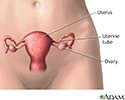
- Uterus
- Vaginal bleeding during pre...
- Well baby visits
- Yeast infections

 Bookmark


