Multimedia Gallery
Human face formation
You might not be aware of this, but during its early development a fetus looks remarkably like something from the dawn of time.
Here, let’s take a closer look. There’s a human fetus’s head during the first month of development, when it was still an embryo. Its face starts as a series of paired tissue mounds called branchial arches.
Let’s take a look from the front. The embryo’s face actually forms from the first branchial arch, along with the area just above it. The forehead and nose form from this area. These areas will form the cheekbones, and these lower areas will form the lower jaw. And this area will form the mouth. At 28 days of development, you can see the lower jaw, which has fused together from the branchial arches. The thickenings you see here will eventually form the nostrils.
By day 31, you can see the nostrils have started to form. And, quite remarkably, the eyes have now appeared on each side of the head.
Two days later, the nostrils have moved toward the center of the face. You can also see that as the ears begin to form, they are positioned in a pretty odd location. But don’t worry, they will move.
At 35 days, the nostrils are even closer together, and we can see more of the eyes. At 40 days, the baby has developed eyelids, and the nose looks much more developed.
Here he is at 48 days and he’s looking pretty darn good. The nasal swellings have joined in the center of the face, and the eyes have moved to the front of the head.
Three weeks later, the fetus looks more human than ever. After that, its face continues to develop more typical proportions right up until the time of its birth.
Let’s look at the entire process again...
As you can see, the development of the face is a fascinating process that has some very dramatic changes taking place in a relatively short amount of time.
Human face formation
Review Date: 8/23/2023
Reviewed By: LaQuita Martinez, MD, Department of Obstetrics and Gynecology, Emory Johns Creek Hospital, Alpharetta, GA. Also reviewed by David C. Dugdale, MD, Medical Director, Brenda Conaway, Editorial Director, and the A.D.A.M. Editorial team.
Animations
- Breast engorgement
- Cell division
- Cesarean section
- Conception - general
- Conception - pregnancy
- Conception of identical twins
- C-section
- Early labor
- Egg cell production
- Egg production
- Endometriosis
- Fetal ear development
- Formation of twins
- Human face formation
- Infant formulas
- Kids - How big is the baby?
- Kids - How does the baby co...
- Kids - Is it a girl or boy?
- Kids - Umbilical cord
- Kids - Where do babies come...
- Newborn jaundice
- NICU consultants and suppor...
- Ovulation
- Placenta delivery
- Placenta formation
- Preeclampsia
- Pregnancy
- Pregnancy care
- Sperm production
- Sperm release pathway
- Storing breast milk
- The role of amniotic fluid
- Twin-to-twin transfusion sy...
- Ultrasound
- Vaginal delivery
Illustrations
- 24-week fetus
- Abnormal discharge from the...
- Abnormal menstrual periods
- Absence of menstruation (am...
- Amniocentesis
- Amniocentesis
- Amniotic fluid
- Amniotic fluid
- Anatomy of a normal placenta
- Antibodies
- Baby burping position
- Bananas and nausea
- Blood cells
- Blood test
- Breast infection
- Breastfeeding
- Bulging fontanelles
- Candida - fluorescent stain
- Caput succedaneum
- Cesarean section
- Cesarean section
- Cesarean section
- Childbirth
- Chorionic villus sampling
- Congenital hip dislocation
- Congenital toxoplasmosis
- Crying - excessive (0 to 6 ...
- Delivery presentations
- Developmental milestones
- Early weeks of pregnancy
- Ectopic pregnancy
- Emergency Childbirth
- Emergency Childbirth
- Endocrine glands
- Endometriosis
- Endometritis
- Erythroblastosis fetalis - ...
- Female breast
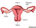
- Female reproductive anatomy
- Female reproductive anatomy
- Female reproductive anatomy...
- Female urinary tract
- Fetal blood testing
- Fetal head molding
- Fetus at 10 weeks
- Fetus at 12 weeks
- Fetus at 16 weeks
- Fetus at 26 to 30 weeks
- Fetus at 3.5 weeks
- Fetus at 30 to 32 weeks
- Fetus at 7.5 weeks
- Fetus at 8.5 weeks
- First trimester of pregnancy
- Folic acid
- Folic acid benefits
- Folic acid source
- Follicle development
- Fontanelles
- Foreskin
- Gestational ages
- Gestational diabetes
- Gonadotropins
- Head circumference
- Heat rash
- Height/weight chart
- Hormonal effects in newborns
- Humidifiers and health
- Hysterectomy
- Infant blood sample
- Infant care following delivery
- Infant diaphragmatic hernia
- Infant heat rash
- Infant intestines
- Infant jaundice
- Infantile reflexes
- Influenza vaccines
- Intraductal papilloma
- Intrauterine transfusion
- Jaundiced infant
- Large fontanelles
- Large fontanelles (lateral view)
- Macrosomia
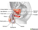
- Male reproductive anatomy
- Male reproductive anatomy
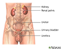
- Male urinary tract
- Mammary gland
- Meconium
- Morning sickness
- Moro reflex
- Newborn head molding
- Newborn test
- Normal female breast anatomy
- Normal uterine anatomy (cut...
- Ovarian cyst
- Ovarian hypofunction
- Overproductive ovaries
- Pelvic adhesions
- Pelvic laparoscopy
- Placenta
- Placenta
- Placenta
- Placenta previa
- Polyhydramnios
- Preeclampsia
- Pregnancy test
- Primary amenorrhea
- Primary infertility
- Secondary amenorrhea
- Secondary infection
- Side sectional view of fema...
- Single palmar crease
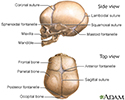
- Skull of a newborn
- Slit-lamp exam
- Sperm
- Stein-Leventhal syndrome
- Sunken fontanelles (superio...
- Tobacco health risks
- Transvaginal ultrasound
- Ultrasound in pregnancy
- Ultrasound, color - normal ...
- Ultrasound, normal fetus - ...
- Ultrasound, normal fetus - ...
- Ultrasound, normal fetus - ...
- Ultrasound, normal fetus - face
- Ultrasound, normal fetus - ...
- Ultrasound, normal fetus - foot
- Ultrasound, normal fetus - ...
- Ultrasound, normal fetus - ...
- Ultrasound, normal fetus - ...
- Ultrasound, normal fetus - ...
- Ultrasound, normal placenta...
- Ultrasound, normal relaxed ...
- Umbilical cord healing
- Uterus
- Vaginal bleeding during pre...
- Well baby visits
- Yeast infections

 Bookmark


