Mediastinitis
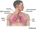
Chest infectionMediastinitis is swelling and irritation (inflammation) of the chest area between the lungs (mediastinum). This area contains the heart, large blood vessels, windpipe (trachea), food tube (esophagus), thymus gland, lymph nodes, and connective tissue.
Causes
Mediastinitis usually results from an infection. It may occur suddenly (acute), or it may develop slowly and get worse over time (chronic). It most often occurs in people who recently had an upper gastrointestinal (GI) endoscopy or chest surgery.
Acute
Acute means sudden. Acute symptoms appear, change, or worsen rapidly. It is the opposite of chronic.

Chronic
Chronic refers to something that continues over an extended period of time. A chronic condition is usually long-lasting and does not easily or quick...

A person may have a tear in their esophagus that causes mediastinitis. Causes of the tear include:
- A procedure such as upper GI endoscopy
Endoscopy
Endoscopy is a way of looking inside the body using a flexible tube that has a small camera and light on the end of it. This instrument is called an...
 ImageRead Article Now Book Mark Article
 - Forceful or constant vomiting
Vomiting
Nausea is feeling an urge to vomit. It is often called "being sick to your stomach. "Vomiting or throwing-up forces the contents of the stomach up t...
 ImageRead Article Now Book Mark Article
 - Trauma
Other causes of mediastinitis include:
- A fungal infection called histoplasmosis
Histoplasmosis
Histoplasmosis is an infection that occurs from breathing in the spores of the fungus Histoplasma capsulatum.
 ImageRead Article Now Book Mark Article
 -
Radiation to the chest
Radiation
Radiation therapy uses high-powered radiation (such as x-rays or gamma rays), particles, or radioactive seeds to kill cancer cells.
 ImageRead Article Now Book Mark Article
 - Inflammation of the lymph nodes, lungs, liver, eyes, skin, or other tissues (sarcoidosis)
Sarcoidosis
Sarcoidosis is a disease in which inflammation occurs in the lymph nodes, lungs, liver, eyes, skin, and other tissues.
 ImageRead Article Now Book Mark Article
 -
Tuberculosis
Tuberculosis
Pulmonary tuberculosis (TB) is a contagious bacterial infection that involves the lungs. It may spread to other organs.
 ImageRead Article Now Book Mark Article
 - Breathing in anthrax
-
Cancer
Cancer
Cancer is the uncontrolled growth of abnormal cells in the body. Cancerous cells are also called malignant cells.
 ImageRead Article Now Book Mark Article
Risk factors include:
- Disease of the esophagus
- Diabetes mellitus
- Problems in the upper gastrointestinal tract
- Recent chest surgery or upper GI endoscopy
- Weakened immune system
Symptoms
Symptoms may include:
-
Chest pain
Chest pain
Chest pain is discomfort or pain that you feel anywhere along the front of your body between your neck and upper abdomen.
 ImageRead Article Now Book Mark Article
 - Chills
- Fever
- General discomfort
-
Shortness of breath
Shortness of breath
Breathing difficulty may involve:Difficult breathing Uncomfortable breathingFeeling like you are not getting enough air
 ImageRead Article Now Book Mark Article
Signs of mediastinitis in people who have had recent surgery include:
- Chest wall tenderness
- Wound drainage
- Unstable chest wall
Exams and Tests
Your health care provider will perform a physical exam and ask about your symptoms and medical history.
Tests may include:
-
Chest CT scan or MRI scan
Chest CT scan
A chest CT (computed tomography) scan is an imaging method that uses x-rays to create cross-sectional pictures of the chest and upper abdomen....
 ImageRead Article Now Book Mark Article
MRI scan
A chest MRI (magnetic resonance imaging) scan is an imaging test that uses powerful magnetic fields and radio waves to create pictures of the chest (...
 ImageRead Article Now Book Mark Article
 -
Chest x-ray
Chest x-ray
A chest x-ray is an x-ray of the chest, lungs, heart, large arteries, ribs, and diaphragm.
 ImageRead Article Now Book Mark Article
 -
Ultrasound
Ultrasound
Ultrasound uses high-frequency sound waves to make images of organs and structures inside the body.
 ImageRead Article Now Book Mark Article
Your provider may insert a needle into the area of inflammation. This is to obtain a sample to send for a Gram stain, other stains and culture to determine the type of infection, if present.
Gram stain
Pleural fluid Gram stain is a method of staining a sample of fluid taken from the pericardium to diagnose a bacterial infection. This is the space s...

Culture
Pleural fluid culture is a test that examines a sample of fluid that has been collected in the pleural space to see if you have an infection to help ...

Treatment
You may receive antibiotics if you have an infection.
You may need surgery to remove the area of inflammation if the blood vessels, windpipe, or esophagus is blocked.
Outlook (Prognosis)
How well a person does depends on the cause and severity of the mediastinitis.
Mediastinitis after chest surgery is very serious. There is a risk of dying from this condition.
Possible Complications
Complications include the following:
- Spread of the infection to the bloodstream, blood vessels, bones, heart, or lungs
- Scarring
Scarring can be severe, especially when it is caused by chronic mediastinitis. Scarring can interfere with heart or lung function.
When to Contact a Medical Professional
Contact your provider if you have had open chest surgery and develop:
- Chest pain
- Chills
- Drainage from the wound
- Fever
- Shortness of breath
If you have a lung infection or sarcoidosis and develop any of these symptoms, contact your provider right away.
Prevention
To lessen the risk of developing mediastinitis related to chest surgery, surgical wounds should be kept clean and dry after surgery.
Kept clean and dry
An incision is a cut through the skin made during surgery. It is also called a surgical wound. Some incisions are small. Others are very long. Th...
Treating tuberculosis, sarcoidosis, or other conditions associated with mediastinitis may prevent this complication.
References
Lentz RJ, Loyd JE. Mediastinitis and fibrosing mediastinitis. In: Broaddus VC, Ernst JD, King TE, et al, eds. Murray and Nadel's Textbook of Respiratory Medicine. 7th ed. Philadelphia, PA: Elsevier; 2022:chap 116.
Van Schooneveld TC, Rupp ME. Mediastinitis. In: Bennett JE, Dolin R, Blaser MJ, eds. Mandell, Douglas, and Bennett's Principles and Practice of Infectious Diseases. 9th ed. Philadelphia, PA: Elsevier; 2020:chap 85.
-
Respiratory system - illustration
Air is breathed in through the nasal passageways, travels through the trachea and bronchi to the lungs.
Respiratory system
illustration
-
Mediastinum - illustration
Mediastinoscopy is a procedure in which a lighted instrument (mediastinoscope) is inserted through a neck incision to visually examine the structures in the top of the chest cavity and take tissue samples. This procedure can be used to biopsy lymph nodes surrounding the airway to help diagnose or see how far a particular disease has spread.
Mediastinum
illustration
-
Respiratory system - illustration
Air is breathed in through the nasal passageways, travels through the trachea and bronchi to the lungs.
Respiratory system
illustration
-
Mediastinum - illustration
Mediastinoscopy is a procedure in which a lighted instrument (mediastinoscope) is inserted through a neck incision to visually examine the structures in the top of the chest cavity and take tissue samples. This procedure can be used to biopsy lymph nodes surrounding the airway to help diagnose or see how far a particular disease has spread.
Mediastinum
illustration
Review Date: 8/29/2024
Reviewed By: Jatin M. Vyas, MD, PhD, Roy and Diana Vagelos Professor in Medicine, Columbia University Vagelos College of Physicians and Surgeons, Division of Infectious Diseases, Department of Medicine, New York, NY. Also reviewed by David C. Dugdale, MD, Medical Director, Brenda Conaway, Editorial Director, and the A.D.A.M. Editorial team.


