Aspiration pneumonia
Anaerobic pneumonia; Aspiration of vomitus; Necrotizing pneumonia; Aspiration pneumonitisPneumonia is inflammation (swelling) and infection of the lungs or large airways.
Aspiration pneumonia occurs when food or liquid is breathed into the airways or lungs, instead of being swallowed.
Causes
Risk factors for breathing in (aspiration) of foreign material into the lungs are:
Aspiration
Aspiration means to draw in or out using a sucking motion. It has two meanings:Breathing in a foreign object (for example, sucking food into the air...

- Being less alert due to medicines, illness, surgery, or other reasons
-
Coma
Coma
Decreased alertness is a state of reduced awareness and is often a serious condition. A coma is the most severe state of decreased alertness in which...
Read Article Now Book Mark Article - Drinking large amounts of alcohol
- Taking illicit drugs (such as opioids) which make you less alert
- Receiving medicine to put you into a deep sleep for surgery (general anesthesia)
General anesthesia
General anesthesia is treatment with certain medicines that puts you into a deep sleep-like state so you do not feel pain during surgery. After you ...
Read Article Now Book Mark Article - Old age
- Poor gag reflex in people who are not alert (unconscious or semi-conscious) after a stroke or brain injury
-
Problems with swallowing
Problems with swallowing
Difficulty with swallowing is the feeling that food or liquid is stuck in the throat or at any point before the food enters the stomach. This proble...
 ImageRead Article Now Book Mark Article
 - Eating or being fed when not upright
Being hospitalized can increase the risk for this condition.
Materials that may be breathed into the lungs include:
- Saliva
- Vomit
- Liquids
- Foods
The type of bacteria that causes the pneumonia depends on:
- Your health
- Where you live (at home or in a long-term nursing facility, for example)
- Whether you were recently hospitalized
- Your recent antibiotic use
- Whether your immune system is weakened
Symptoms
Symptoms may include any of the following:
- Chest pain
- Coughing up foul-smelling, greenish or dark phlegm (sputum), or phlegm that contains pus or blood
-
Fatigue
Fatigue
Fatigue is a feeling of weariness, tiredness, or lack of energy.
 ImageRead Article Now Book Mark Article
 - Fever
-
Shortness of breath
Shortness of breath
Breathing difficulty may involve:Difficult breathing Uncomfortable breathingFeeling like you are not getting enough air
 ImageRead Article Now Book Mark Article
 -
Wheezing
Wheezing
Wheezing is a high-pitched whistling sound during breathing. It occurs when air moves through narrowed breathing tubes in the lungs.
 ImageRead Article Now Book Mark Article
 -
Breath odor
Breath odor
Breath odor is the scent of the air you breathe out of your mouth. Unpleasant breath odor is commonly called bad breath.
 ImageRead Article Now Book Mark Article
 - Excessive sweating
- Problems swallowing
- Confusion
- Seeing food or tube feed material (if being fed artificially) in your sputum
Exams and Tests
Your health care provider will use a stethoscope to listen for crackles or abnormal breath sounds in your chest. Tapping on your chest wall (percussion) helps the provider listen and feel for abnormal sounds in your chest.
If pneumonia is suspected, your provider will likely order a chest x-ray.
Chest x-ray
A chest x-ray is an x-ray of the chest, lungs, heart, large arteries, ribs, and diaphragm.

The following tests also may help diagnose this condition:
-
Arterial blood gas
Arterial blood gas
Blood gases are a measurement of how much oxygen and carbon dioxide are in your blood. They also determine the acidity (pH) of your blood.
 ImageRead Article Now Book Mark Article
 -
Blood culture
Blood culture
A blood culture is a laboratory test to check for bacteria or other germs in a blood sample.
 ImageRead Article Now Book Mark Article
 -
Bronchoscopy (uses a special scope to view the lung airways) in some cases
Bronchoscopy
Bronchoscopy is a test to view the airways and diagnose lung disease. It may also be used during the treatment of some lung conditions.
 ImageRead Article Now Book Mark Article
 - Complete blood count (CBC)
CBC
A complete blood count (CBC) test measures the following:The number of white blood cells (WBC count)The number of red blood cells (RBC count)The numb...
 ImageRead Article Now Book Mark Article
 -
X-rays or CT scan of the chest
CT scan
A computed tomography (CT) scan is an imaging method that uses x-rays to create pictures of cross-sections of the body. Related tests include:Abdomin...
 ImageRead Article Now Book Mark Article
 -
Sputum culture
Sputum culture
Routine sputum culture is a laboratory test that looks for germs that cause infection. Sputum is the material that comes up from air passages when y...
 ImageRead Article Now Book Mark Article
 - Swallowing tests
Treatment
Some people may need to be hospitalized. Treatment depends on how severe the pneumonia is and how ill the person is before the aspiration (chronic illness). Sometimes a ventilator (breathing machine) is needed to support breathing.
Ventilator (breathing machine)
A ventilator is a machine that breathes for you or helps you breathe. It is also called a breathing machine or respirator. The ventilator: Is a com...
You will likely receive antibiotics.
You may need to have your swallowing function tested. People who have trouble swallowing may need to use other feeding methods to reduce the risk of aspiration.
Outlook (Prognosis)
Outcome depends on:
- The health of the person before getting pneumonia
- The type of bacteria causing the pneumonia
- How much of the lungs are involved
More severe infections may result in long-term damage to the lungs.
Possible Complications
Complications may include:
- Lung abscess
Abscess
An abscess is a collection of pus in any part of the body. In most cases, the area around an abscess is swollen and inflamed.
 ImageRead Article Now Book Mark Article
 -
Shock
Shock
Shock is a life-threatening condition that occurs when the body is not getting enough blood flow. Lack of blood flow means the cells and organs do n...
 ImageRead Article Now Book Mark Article
 - Spread of infection to the bloodstream (bacteremia)
Bacteremia
Septicemia is an infection in the bloodstream that is caused by bacteria, viruses, or fungi. Also called sepsis, septicemia is a serious, life-threa...
Read Article Now Book Mark Article - Spread of infection to other areas of the body
- Respiratory failure
- Death
When to Contact a Medical Professional
Contact your provider, go to the emergency room, or call the local emergency number (such as 911) if you have:
- Chest pain
- Chills
- Fever
- Shortness of breath
- Bluish discoloration of the lips or tongue (cyanosis)
- Wheezing
References
Baden LR, Griffin MR, Klompas M. Overview of pneumonia. In: Goldman L, Cooney KA, eds. Goldman-Cecil Medicine. 27th ed. Philadelphia, PA: Elsevier; 2024:chap 85.
Shah RJ, Young VN. Aspiration. In: Broaddus VC, Ernst JD, King TE, et al, eds. Murray and Nadel's Textbook of Respiratory Medicine. 7th ed. Philadelphia, PA: Elsevier; 2022:chap 43.
-
Pneumococci organism - illustration
This picture shows the organism Pneumococci. These bacteria are usually paired (diplococci) or appear in chains. Pneumococci are typically associated with pneumonia, but may cause infection in other organs such as the brain (pneumococcal meningitis) and blood stream (pneumococcal septicemia). (Image courtesy of the Centers for Disease Control and Prevention)
Pneumococci organism
illustration
-
Bronchoscopy - illustration
Bronchoscopy is a surgical technique for viewing the interior of the airways. Using sophisticated flexible fiber optic instruments, surgeons are able to explore the trachea, main stem bronchi, and some of the small bronchi. In children, this procedure may be used to remove foreign objects that have been inhaled. In adults, the procedure is most often used to take samples of (biopsy) suspicious lesions and for culturing specific areas in the lung.
Bronchoscopy
illustration
-
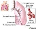
Lungs - illustration
The major features of the lungs include the bronchi, the bronchioles and the alveoli. The alveoli are the microscopic blood vessel-lined sacks in which oxygen and carbon dioxide gas are exchanged.
Lungs
illustration
-
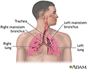
Respiratory system - illustration
Air is breathed in through the nasal passageways, travels through the trachea and bronchi to the lungs.
Respiratory system
illustration
-
Pneumococci organism - illustration
This picture shows the organism Pneumococci. These bacteria are usually paired (diplococci) or appear in chains. Pneumococci are typically associated with pneumonia, but may cause infection in other organs such as the brain (pneumococcal meningitis) and blood stream (pneumococcal septicemia). (Image courtesy of the Centers for Disease Control and Prevention)
Pneumococci organism
illustration
-
Bronchoscopy - illustration
Bronchoscopy is a surgical technique for viewing the interior of the airways. Using sophisticated flexible fiber optic instruments, surgeons are able to explore the trachea, main stem bronchi, and some of the small bronchi. In children, this procedure may be used to remove foreign objects that have been inhaled. In adults, the procedure is most often used to take samples of (biopsy) suspicious lesions and for culturing specific areas in the lung.
Bronchoscopy
illustration
-
Lungs - illustration
The major features of the lungs include the bronchi, the bronchioles and the alveoli. The alveoli are the microscopic blood vessel-lined sacks in which oxygen and carbon dioxide gas are exchanged.
Lungs
illustration
-
Respiratory system - illustration
Air is breathed in through the nasal passageways, travels through the trachea and bronchi to the lungs.
Respiratory system
illustration
Review Date: 8/13/2023
Reviewed By: Denis Hadjiliadis, MD, MHS, Paul F. Harron, Jr. Professor of Medicine, Pulmonary, Allergy, and Critical Care, Perelman School of Medicine, University of Pennsylvania, Philadelphia, PA. Also reviewed by David C. Dugdale, MD, Medical Director, Brenda Conaway, Editorial Director, and the A.D.A.M. Editorial team.


