Pulmonary edema
Lung congestion; Lung water; Pulmonary congestion; Heart failure - pulmonary edemaPulmonary edema is an abnormal buildup of fluid in the lungs. This buildup of fluid leads to shortness of breath.
Causes
Pulmonary edema is often caused by congestive heart failure. When the heart is not able to pump efficiently, blood can back up into the blood vessels that take blood through the lungs.
Congestive heart failure
Heart failure is a condition in which the heart is no longer able to pump oxygen-rich blood to the rest of the body efficiently. This causes symptom...

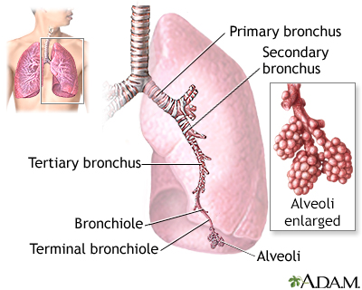
As the pressure in these blood vessels increases, fluid is pushed into the air spaces (alveoli) in the lungs. This fluid reduces normal oxygen movement through the lungs. These two factors combine to cause shortness of breath.
Congestive heart failure that leads to pulmonary edema may be caused by:
- Heart attack, or any disease of the heart that weakens or stiffens the heart muscle (cardiomyopathy)
Cardiomyopathy
Cardiomyopathy is disease in which the heart muscle becomes weakened, stretched, or has another structural problem. It often contributes to the hear...
 ImageRead Article Now Book Mark Article
 - Leaking or narrowed heart valves (mitral or aortic valves)
- Sudden, severe high blood pressure (hypertension)
Pulmonary edema may also be caused by:
- Certain medicines
- High altitude exposure
- Kidney failure
- Narrowed arteries that bring blood to the kidneys
- Lung damage caused by burns from poisonous gas inhalation or severe infection
- Major injury
Symptoms
Symptoms of pulmonary edema may include:
- Coughing up blood or bloody froth
- Difficulty breathing when lying down (orthopnea)
- Feeling of "air hunger" or "drowning" (This feeling is called "paroxysmal nocturnal dyspnea" if it causes you to wake up 1 to 2 hours after falling asleep and struggle to catch your breath.)
- Grunting, gurgling, or wheezing sounds with breathing
- Problems speaking in full sentences because of shortness of breath
Other symptoms may include:
- Anxiety or restlessness
- Decrease in level of alertness
- Leg or abdominal swelling
- Pale skin
- Sweating (excessive)
Exams and Tests
The health care provider will perform a thorough physical exam.
The provider will listen to your lungs and heart with a stethoscope to check for:
-
Abnormal heart sounds
Abnormal heart sounds
A heart murmur is a blowing, whooshing, or rasping sound heard during a heartbeat. The sound is caused by turbulent (rough) blood flow through the h...
 ImageRead Article Now Book Mark Article
 - Crackles in your lungs, called rales
- Increased heart rate (tachycardia)
- Rapid breathing (tachypnea)
Other things that may be seen during the exam include:
- Leg or abdominal swelling
- Abnormalities of your neck veins (which can show that there is too much fluid in your body)
- Pale or blue skin color (pallor or cyanosis)
Possible tests include:
- Blood chemistries
- Blood oxygen levels (oximetry or arterial blood gases)
-
Chest x-ray
Chest x-ray
A chest x-ray is an x-ray of the chest, lungs, heart, large arteries, ribs, and diaphragm.
 ImageRead Article Now Book Mark Article
 - Complete blood count (CBC)
-
Echocardiogram (ultrasound of the heart) to see if there are problems with the heart muscle
Echocardiogram
An echocardiogram is a test that uses sound waves to create pictures of the heart. The picture and information it produces is more detailed than a s...
 ImageRead Article Now Book Mark Article
 -
Electrocardiogram (ECG) to look for signs of a heart attack or problems with the heart rhythm
Electrocardiogram
An electrocardiogram (ECG) is a test that records the electrical activity of the heart.
 ImageRead Article Now Book Mark Article
Treatment
Pulmonary edema is almost always treated in the emergency room or hospital. You may need to be in an intensive care unit (ICU).
- Oxygen is given through a face mask or tiny plastic tubes are placed in the nose.
- A breathing tube may be placed into the windpipe (trachea) so you can be connected to a breathing machine (ventilator) if you cannot breathe well on your own (only needed in severe cases).
The cause of pulmonary edema should be identified and treated quickly. For example, if a heart attack has caused the condition, it must be treated right away.
Medicines that may be used include:
- Diuretics that remove excess fluid from the body
- Medicines that strengthen the heart muscle, control the heartbeat, or relieve pressure on the heart
- Other medicines when heart failure is not the cause of the pulmonary edema
Outlook (Prognosis)
The outlook depends on the cause. The condition may get better quickly or slowly. Some people may need to use a breathing machine for a long time. If not treated, this condition can be life threatening.
When to Contact a Medical Professional
Go to the emergency room or call 911 or the local emergency number if you have breathing problems.
Prevention
Take all your medicines as directed if you have a disease that can lead to pulmonary edema or a weakened heart muscle.
Following a healthy diet that is low in salt and fat, and controlling your other risk factors can reduce the risk of developing this condition.
References
Mann DL. Management of heart failure patients with reduced ejection fraction. In: Libby P, Bonow RO, Mann DL, Tomaselli GF, Bhatt DL, Solomon SD, eds. Braunwald's Heart Disease: A Textbook of Cardiovascular Medicine. 12th ed. Philadelphia, PA: Elsevier; 2022:chap 50.
Meyer NJ, Matthay MA. Pulmonary edema. In: Broaddus VC, Ernst JD, King TE, et al, eds. Murray and Nadel's Textbook of Respiratory Medicine. 7th ed. Philadelphia, PA: Elsevier; 2022:chap 133.
Rogers JG, O'Connor CM. Heart failure: epidemiology, pathobiology, and diagnosis. In: Goldman L, Cooney KA, eds. Goldman-Cecil Medicine. 27th ed. Philadelphia, PA: Elsevier; 2024:chap 45.
-
Congestive heart failure (CHF) overview
Animation
-
Lungs - illustration
The major features of the lungs include the bronchi, the bronchioles and the alveoli. The alveoli are the microscopic blood vessel-lined sacks in which oxygen and carbon dioxide gas are exchanged.
Lungs
illustration
-
Respiratory system - illustration
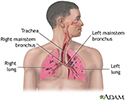
Air is breathed in through the nasal passageways, travels through the trachea and bronchi to the lungs.
Respiratory system
illustration
-
Lungs - illustration
The major features of the lungs include the bronchi, the bronchioles and the alveoli. The alveoli are the microscopic blood vessel-lined sacks in which oxygen and carbon dioxide gas are exchanged.
Lungs
illustration
-
Respiratory system - illustration
Air is breathed in through the nasal passageways, travels through the trachea and bronchi to the lungs.
Respiratory system
illustration
Review Date: 2/27/2024
Reviewed By: Thomas S. Metkus, MD, Assistant Professor of Medicine and Surgery, Johns Hopkins University School of Medicine, Baltimore, MD. Also reviewed by David C. Dugdale, MD, Medical Director, Brenda Conaway, Editorial Director, and the A.D.A.M. Editorial team.


