Cardiac amyloidosis
Amyloidosis - cardiac; Primary cardiac amyloidosis - AL type; Secondary cardiac amyloidosis - AA type; Stiff heart syndrome; Senile amyloidosisCardiac amyloidosis is a disorder caused by deposits of an abnormal protein (amyloid) in the heart tissue. These deposits make it hard for the heart to work properly.
Causes
Amyloidosis is a group of diseases in which clumps of proteins called amyloid build up in body tissues. Over time, these proteins replace normal tissue, leading to failure of the involved organ. There are many forms of amyloidosis.
Amyloidosis
Primary amyloidosis is a rare disorder in which abnormal proteins build up in tissues and organs. Clumps of the abnormal proteins are called amyloid...

Cardiac amyloidosis ("stiff heart syndrome") occurs when amyloid deposits take the place of normal heart muscle. It is the most typical type of restrictive cardiomyopathy. Cardiac amyloidosis may affect the way electrical signals move through the heart (conduction system). This can lead to abnormal heartbeats (arrhythmias) and faulty heart signals (heart block).
Restrictive cardiomyopathy
Restrictive cardiomyopathy refers to a set of changes in how the heart muscle functions. These changes cause the heart to fill poorly (more common) ...

Arrhythmias
An arrhythmia is a disorder of the heart rate (pulse) or heart rhythm. The heart can beat too fast (tachycardia), too slow (bradycardia), or irregul...

The condition can be inherited. This is called familial cardiac amyloidosis. It can also develop as the result of another disease such as a type of bone and blood cancer, or as the result of another medical problem causing inflammation. Cardiac amyloidosis is more common in men than in women. The disease is rare in people under age 40.
Symptoms
Some people may have no symptoms. When present, symptoms may include:
- Excessive urination at night
- Fatigue, reduced exercise ability
- Palpitations (sensation of feeling heartbeat)
- Shortness of breath with activity
- Swelling of the abdomen, legs, ankles, or other part of the body
- Trouble breathing while lying down
Exams and Tests
The signs of cardiac amyloidosis can be related to a number of different conditions. This can make the problem hard to diagnose.
Signs may include:
- Abnormal sounds in the lung (lung crackles) or a heart murmur
- Blood pressure that is low or drops when you stand up
- Enlarged neck veins
- Swollen liver
The following tests may be done:
- Blood and urine protein tests
-
Chest or abdomen CT scan (considered the "gold standard" to help diagnose this condition)
Chest or abdomen CT scan
A chest CT (computed tomography) scan is an imaging method that uses x-rays to create cross-sectional pictures of the chest and upper abdomen....
 ImageRead Article Now Book Mark Article
 -
Coronary angiography
Coronary angiography
Coronary angiography is a procedure that uses a special dye (contrast material) and x-rays to see how blood flows through the arteries in your heart....
 ImageRead Article Now Book Mark Article
 -
Echocardiogram
Echocardiogram
An echocardiogram is a test that uses sound waves to create pictures of the heart. The picture and information it produces is more detailed than a s...
 ImageRead Article Now Book Mark Article
 -
Electrocardiogram (ECG)
Electrocardiogram (ECG)
An electrocardiogram (ECG) is a test that records the electrical activity of the heart.
 ImageRead Article Now Book Mark Article
 -
Magnetic resonance imaging (MRI)
Magnetic resonance imaging
A magnetic resonance imaging (MRI) scan is an imaging test that uses powerful magnets and radio waves to create pictures of the body. It does not us...
 ImageRead Article Now Book Mark Article
 -
Nuclear heart scans (MUGA, RNV)
Nuclear heart scans
Nuclear ventriculography is a test that uses radioactive materials called tracers to show the heart chambers. The procedure is noninvasive. The ins...
 ImageRead Article Now Book Mark Article
 - Positron emission tomography (PET)
An ECG may show problems with the heartbeat or heart signals. It may also show low signals (called "low voltage").
A cardiac biopsy may be used to confirm the diagnosis. A biopsy of another area, such as the abdomen, kidney, or bone marrow, is often done as well.
Cardiac biopsy
Myocardial biopsy is the removal of a small piece of heart muscle for examination.

Treatment
Your health care provider may tell you to make changes to your diet, including limiting salt and fluids.
You may need to take water pills (diuretics) to help your body get rid of excess fluid. Your provider may tell you to weigh yourself every day. A weight gain of 3 or more pounds (1 kilogram or more) over 1 to 2 days can mean there is too much fluid in the body.
Medicines including digoxin, calcium-channel blockers, and beta-blockers may be used in people with atrial fibrillation. However, the medicines must be used with caution, and the dosage must be carefully monitored. People with cardiac amyloidosis may be extra sensitive to side effects of these medicines.
Other treatments may include:
- Chemotherapy
- Drugs that target the abnormal protein (tafamidis)
- Implantable cardioverter-defibrillator (AICD)
- Pacemaker, if there are problems with heart signals
- Prednisone, an anti-inflammatory medicine
A heart transplant may be considered for people with some types of amyloidosis who have very poor heart function. People with hereditary amyloidosis may need a liver transplant.
Heart transplant
A heart transplant is surgery to remove a damaged or diseased heart and replace it with a healthy donor heart.

Outlook (Prognosis)
In the past, cardiac amyloidosis was thought to be an untreatable and rapidly fatal disease. However, the field is changing rapidly. Different types of amyloidosis can affect the heart in different ways. Some types are more severe than others. Many people can now expect to survive and experience a good quality of life for several years after diagnosis.
Possible Complications
Complications may include:
-
Atrial fibrillation or ventricular arrhythmias
Atrial fibrillation
Atrial fibrillation (AFib) and atrial flutter are common types of abnormal heart rhythms (arrhythmias) which affect the upper chambers (atria) of the...
 ImageRead Article Now Book Mark Article
 -
Congestive heart failure
Congestive heart failure
Heart failure is a condition in which the heart is no longer able to pump oxygen-rich blood to the rest of the body efficiently. This causes symptom...
 ImageRead Article Now Book Mark Article
 - Fluid buildup in the abdomen (ascites)
Ascites
Ascites is the build-up of fluid in the space between the lining of the abdomen and abdominal organs.
 ImageRead Article Now Book Mark Article
 - Increased sensitivity to digoxin
- Low blood pressure and dizziness from excessive urination (due to medicine)
-
Sick sinus syndrome
Sick sinus syndrome
Normally, the heartbeat starts in an area in the top chambers of the heart (atria). This area is the heart's pacemaker. It is called the sinoatrial...
 ImageRead Article Now Book Mark Article
 -
Symptomatic cardiac conduction system disease (arrhythmias related to abnormal conduction of impulses through the heart muscle)
Symptomatic
Symptomatic can mean showing symptoms, or it may concern a specific symptom. Symptoms may be signs of disease or injury. They are what a person fee...
 ImageRead Article Now Book Mark Article
When to Contact a Medical Professional
Contact your provider if you have this disorder and develop new symptoms such as:
-
Dizziness when you change position
Dizziness
Dizziness is a term that is often used to describe 2 different symptoms: lightheadedness and vertigo. Lightheadedness is a feeling that you might fai...
 ImageRead Article Now Book Mark Article
 - Excessive weight (fluid) gain
- Excessive weight loss
-
Fainting spells
Fainting
Fainting is a brief loss of consciousness due to a drop in blood flow to the brain. The episode most often lasts less than a couple of minutes and y...
Read Article Now Book Mark Article - Severe breathing problems
References
Elliott PM, Olivotto I. Diseases of the myocardium and endocardium. In: Goldman L, Cooney KA, eds. Goldman-Cecil Medicine. 27th ed. Philadelphia, PA: Elsevier; 2024:chap 47.
Hershberger RE. The dilated, restrictive, and infiltrative cardiomyopathies. In: Libby P, Bonow RO, Mann DL, Tomaselli GF, Bhatt DL, Solomon SD, eds. Braunwald's Heart Disease: A Textbook of Cardiovascular Medicine. 12th ed. Philadelphia, PA: Elsevier; 2022:chap 52.
-
Heart - section through the middle - illustration
The interior of the heart is composed of valves, chambers, and associated vessels.
Heart - section through the middle
illustration
-
Dilated cardiomyopathy - illustration
Dilated cardiomyopathy involves enlargement of the heart muscle and is the most common type of cardiomyopathy. The heart muscle is weakened and cannot pump blood efficiently. Decreased heart function affects the lungs, liver, and other body systems.
Dilated cardiomyopathy
illustration
-
Biopsy catheter - illustration
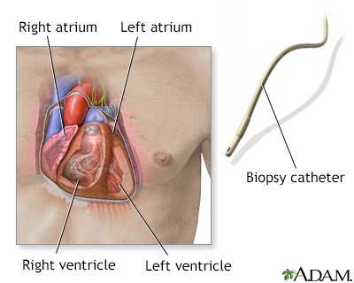
When a small piece of heart muscle tissue is needed for examination, a heart biopsy can be performed. A catheter is carefully threaded into an artery or vein to gain access into the heart. A bioptome (catheter with jaws in its tip) is then introduced. Once the bioptome is in place, three to five small pieces of tissue from the heart muscle are removed. The test is performed routinely after heart transplantation to detect potential rejection. It may also be performed when cardiomyopathy, myocarditis, cardiac amyloidosis, or other disorders are suspected.
Biopsy catheter
illustration
-
Heart - section through the middle - illustration
The interior of the heart is composed of valves, chambers, and associated vessels.
Heart - section through the middle
illustration
-
Dilated cardiomyopathy - illustration
Dilated cardiomyopathy involves enlargement of the heart muscle and is the most common type of cardiomyopathy. The heart muscle is weakened and cannot pump blood efficiently. Decreased heart function affects the lungs, liver, and other body systems.
Dilated cardiomyopathy
illustration
-
Biopsy catheter - illustration
When a small piece of heart muscle tissue is needed for examination, a heart biopsy can be performed. A catheter is carefully threaded into an artery or vein to gain access into the heart. A bioptome (catheter with jaws in its tip) is then introduced. Once the bioptome is in place, three to five small pieces of tissue from the heart muscle are removed. The test is performed routinely after heart transplantation to detect potential rejection. It may also be performed when cardiomyopathy, myocarditis, cardiac amyloidosis, or other disorders are suspected.
Biopsy catheter
illustration
Review Date: 5/8/2024
Reviewed By: Thomas S. Metkus, MD, Assistant Professor of Medicine and Surgery, Johns Hopkins University School of Medicine, Baltimore, MD. Also reviewed by David C. Dugdale, MD, Medical Director, Brenda Conaway, Editorial Director, and the A.D.A.M. Editorial team.


