Chronic cholecystitis
Cholecystitis - chronicChronic cholecystitis is swelling and irritation of the gallbladder that continues over time.
The gallbladder is a sac located under the liver. It stores bile that is made in the liver.
Bile helps with the digestion of fats in the small intestine.
Causes
Most of the time, chronic cholecystitis is caused by repeated attacks of acute (sudden) cholecystitis. Most of these attacks are caused by gallstones in the gallbladder.
These attacks cause the walls of the gallbladder to thicken. The gallbladder begins to shrink. Over time, the gallbladder is less able to concentrate, store, and release bile.
The disease occurs more often in women than in men. It is more common after age 40. Birth control pills and pregnancy are factors that increase the risk for gallstones.
Symptoms
Acute cholecystitis is a painful condition that may lead to chronic cholecystitis. It is not clear whether chronic cholecystitis causes any symptoms.
Acute cholecystitis
Acute cholecystitis is sudden swelling and irritation of the gallbladder. It causes severe belly pain.

Symptoms of acute cholecystitis can include:
- Sharp, cramping, or dull pain in upper right or upper middle of your belly
- Steady pain lasting about 30 minutes
- Pain that spreads to your back or below your right shoulder blade
- Clay-colored stools
- Fever
- Nausea and vomiting
- Yellowing of skin and whites of the eyes (jaundice)
Exams and Tests
Your health care provider may order the following blood tests:
-
Lipase in order to diagnose diseases of the pancreas
Lipase
Lipase is a protein (enzyme) released by the pancreas into the small intestine. It helps the body absorb fat. This test is used to measure the amou...
 ImageRead Article Now Book Mark Article
 -
Complete blood count (CBC)
Complete blood count
A complete blood count (CBC) test measures the following:The number of white blood cells (WBC count)The number of red blood cells (RBC count)The numb...
 ImageRead Article Now Book Mark Article
 -
Liver function tests in order to evaluate how well the liver is working
Liver function tests
Liver function tests are common tests that are used to see how well the liver is working. Tests include:AlbuminAlpha-1 antitrypsinAlkaline phosphata...
 ImageRead Article Now Book Mark Article
Tests that reveal gallstones or inflammation in the gallbladder include:
-
Abdominal ultrasound
Abdominal ultrasound
Abdominal ultrasound is a type of imaging test. It is used to look at organs in the abdomen, including the liver, gallbladder, pancreas, and kidneys...
 ImageRead Article Now Book Mark Article
 -
Abdominal CT scan
Abdominal CT scan
An abdominal CT scan is an imaging test that uses x-rays to create cross-sectional pictures of the belly area. CT stands for computed tomography....
 ImageRead Article Now Book Mark Article
 - Gallbladder scan (HIDA scan)
- Oral cholecystogram (rarely done)
Treatment
Surgery is the most common treatment. Surgery to remove the gallbladder is called cholecystectomy.
- Laparoscopic cholecystectomy is most often done. This surgery uses smaller surgical cuts, which results in a faster recovery. Many people are able to go home from the hospital on the same day as surgery, or the next morning.
- Open cholecystectomy requires a larger cut in the upper-right part of the abdomen.
If you are too ill to have surgery because of other diseases or conditions, the gallstones may be dissolved with medicine you take by mouth. However, this may take 2 years or longer to work. The stones may return after treatment.
Outlook (Prognosis)
Cholecystectomy is a common procedure with a low risk.
Possible Complications
Complications may include:
-
Cancer of the gallbladder (rarely). Sometimes cancer is found in a gallbladder that has been removed.
Cancer
Cancer is the uncontrolled growth of abnormal cells in the body. Cancerous cells are also called malignant cells.
 ImageRead Article Now Book Mark Article
 - Jaundice.
- Pancreatitis.
- Worsening of the condition.
When to Contact a Medical Professional
Contact your provider if you develop symptoms of cholecystitis.
Prevention
The condition is not always preventable. Eating less fatty foods may relieve symptoms in people. However, the benefit of a low-fat diet has not been proven.
References
Basturk O, Adsay NV. Diseases of the gallbladder. In: Burt AD, Ferrell LD, Hubscher SG, eds. MacSween's Pathology of the Liver. 8th ed. Philadelphia, PA: Elsevier; 2024:chap 10.
Gill RM, Kakar S. Liver and gallbladder. In: Kumar V, Abbas AK, Aster JC, eds. Robbins and Cotran Pathologic Basis of Disease. 10th ed. Philadelphia, PA: Elsevier; 2021:chap 18.
Wang DQH, Afdhal NH. Gallstone disease. In: Feldman M, Friedman LS, Brandt LJ, eds. Sleisenger and Fordtran's Gastrointestinal and Liver Disease. 11th ed. Philadelphia, PA: Elsevier; 2021:chap 65.
-
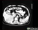
Cholecystitis, CT scan - illustration
This is a CT scan of the upper abdomen showing cholecystitis (gall stones).
Cholecystitis, CT scan
illustration
-
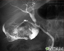
Cholecystitis, cholangiogram - illustration
Cholelithiasis can be seen on a cholangiogram. Radio-opaque dye is used to enhance the x-ray. Multiple stones are present in the gallbladder (PTCA).
Cholecystitis, cholangiogram
illustration
-
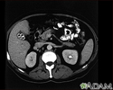
Cholecystolithiasis - illustration
Cholecystolithiasis. CT scan of the upper abdomen showing multiple gallstones.
Cholecystolithiasis
illustration
-
Gallstones, cholangiogram - illustration
A cholecystogram in a patient with gallstones.
Gallstones, cholangiogram
illustration
-
Cholecystitis, CT scan - illustration
This is a CT scan of the upper abdomen showing cholecystitis (gall stones).
Cholecystitis, CT scan
illustration
-
Cholecystitis, cholangiogram - illustration
Cholelithiasis can be seen on a cholangiogram. Radio-opaque dye is used to enhance the x-ray. Multiple stones are present in the gallbladder (PTCA).
Cholecystitis, cholangiogram
illustration
-
Cholecystolithiasis - illustration
Cholecystolithiasis. CT scan of the upper abdomen showing multiple gallstones.
Cholecystolithiasis
illustration
-
Gallstones, cholangiogram - illustration
A cholecystogram in a patient with gallstones.
Gallstones, cholangiogram
illustration
Review Date: 12/31/2023
Reviewed By: Jenifer K. Lehrer, MD, Department of Gastroenterology, Aria - Jefferson Health Torresdale, Jefferson Digestive Diseases Network, Philadelphia, PA. Review provided by VeriMed Healthcare Network. Also reviewed by David C. Dugdale, MD, Medical Director, Brenda Conaway, Editorial Director, and the A.D.A.M. Editorial team.


