Intestinal obstruction and Ileus
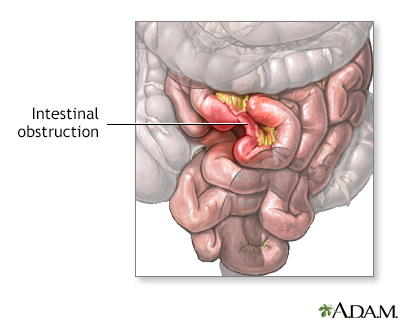
Paralytic ileus; Intestinal volvulus; Bowel obstruction; Ileus; Pseudo-obstruction - intestinal; Colonic ileus; Small bowel obstructionIntestinal obstruction is a partial or complete blockage of the bowel. The contents of the intestine cannot pass through it.
Causes
Obstruction of the bowel may be due to:
- A mechanical cause, which means something is partially of fully blocking the bowel
- Ileus, a condition in which the bowel does not work correctly, but there is no structural problem causing the obstruction
Paralytic ileus, also called pseudo-obstruction, is one of the major causes of intestinal obstruction in infants and children. Causes of paralytic ileus may include:
- Bacteria or viruses that cause intestinal infections (gastroenteritis)
Gastroenteritis
Bacterial gastroenteritis occurs when there is a bacterial infection of your stomach or intestines.
 ImageRead Article Now Book Mark Article
 - Chemical, electrolyte, or mineral imbalances (such as decreased blood potassium level)
- Abdominal surgery
-
Decreased blood supply to the intestines
Decreased blood supply to the intestine
Mesenteric artery ischemia occurs when there is a narrowing or blockage of one or more of the three major arteries that supply the small and large in...
 ImageRead Article Now Book Mark Article
 - Infections inside the abdomen, such as appendicitis
- Kidney or lung disease
- Use of certain medicines, especially narcotics
Mechanical causes of intestinal obstruction may include:
-
Adhesions or scar tissue that form after surgery
Adhesions
Adhesions are bands of scar-like tissue that form between two surfaces inside the body and cause them to stick together.
 ImageRead Article Now Book Mark Article
 - Foreign bodies (objects that are swallowed and block the intestines)
-
Gallstones (rare)
Gallstones
Gallstones are hard deposits that form inside the gallbladder. These may be as small as a grain of sand or as large as a golf ball.
 ImageRead Article Now Book Mark Article
 -
Hernias
Hernias
A hernia is a sac formed by the lining of the abdominal cavity (peritoneum). The sac comes through a hole or weak area in the strong layer of the be...
 ImageRead Article Now Book Mark Article
 -
Impacted stool
Impacted stool
A fecal impaction is a large lump of dry, hard stool that stays stuck in the rectum. It is most often seen in people who are constipated for a long ...
 ImageRead Article Now Book Mark Article
 -
Intussusception (telescoping of one segment of bowel into another)
Intussusception
Intussusception is the sliding of one part of the intestine into another. This article focuses on intussusception in children.
 ImageRead Article Now Book Mark Article
 -
Tumors blocking the intestines
Tumors
A tumor is an abnormal growth of body tissue. Tumors can be cancerous (malignant) or noncancerous (benign).
Read Article Now Book Mark Article - Volvulus (twisted intestine)
- Inflammatory diseases such as Crohn disease
Symptoms
Symptoms may include:
- Abdominal swelling (distention)
-
Abdominal fullness, gas
Abdominal fullness, gas
Gas is air in the intestine that is passed through the rectum. Air that moves from the digestive tract through the mouth is called belching. Gas is ...
 ImageRead Article Now Book Mark Article
 -
Abdominal pain and cramping
Abdominal pain
Abdominal pain is pain that you feel anywhere between your chest and groin. This is often referred to as the stomach region or belly.
 ImageRead Article Now Book Mark Article
 -
Breath odor
Breath odor
Breath odor is the scent of the air you breathe out of your mouth. Unpleasant breath odor is commonly called bad breath.
 ImageRead Article Now Book Mark Article
 -
Constipation
Constipation
Constipation in infants and children means they have hard stools or have problems passing stools. A child may have pain while passing stools or may ...
 ImageRead Article Now Book Mark Article
 - Diarrhea
- Inability to pass gas
-
Nausea and vomiting
Vomiting
Nausea is feeling an urge to vomit. It is often called "being sick to your stomach. "Vomiting or throwing-up forces the contents of the stomach up t...
 ImageRead Article Now Book Mark Article
Exams and Tests
During a physical exam, the health care provider may find bloating, tenderness, or hernias in the abdomen.
Hernias
A hernia is a sac formed by the lining of the abdominal cavity (peritoneum). The sac comes through a hole or weak area in the strong layer of the be...

Tests that show obstruction include:
- Abdominal CT scan
CT scan
A computed tomography (CT) scan is an imaging method that uses x-rays to create pictures of cross-sections of the body. Related tests include:Abdomin...
 ImageRead Article Now Book Mark Article
 - Abdominal x-ray
-
Barium enema
Barium enema
A barium enema is a special x-ray of the large intestine, which includes the colon and rectum.
 ImageRead Article Now Book Mark Article
 -
Upper GI and small bowel series
Upper GI and small bowel series
An upper GI and small bowel series is a set of x-rays taken to examine the esophagus, stomach, and small intestine. Barium enema is a different test ...
 ImageRead Article Now Book Mark Article
Treatment
Treatment involves placing a tube through the nose into the stomach or intestine. This is to help relieve abdominal swelling (distention) and vomiting. Volvulus of the large bowel may be treated by passing a tube into the rectum.
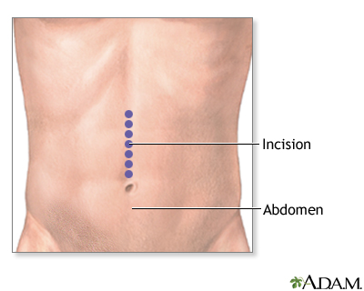
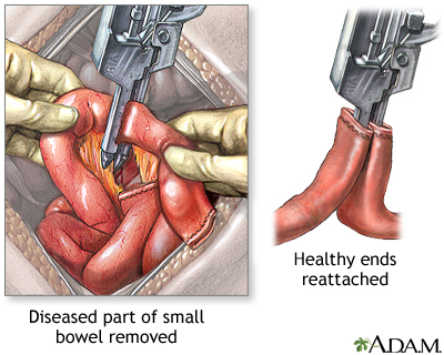
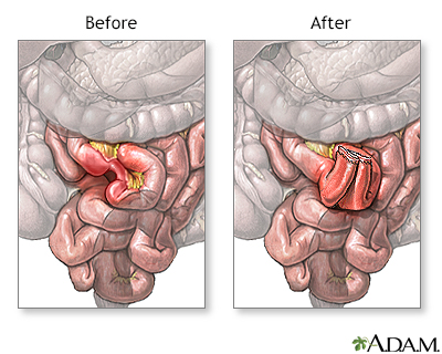
Surgery may be needed to relieve the obstruction if the tube does not relieve the symptoms. It may also be needed if there are signs of tissue death. If a tumor is causing a mechanical obstruction, surgery may be needed. Obstruction from inflammation such as Crohn disease may be treated with surgery, a procedure to dilate the narrow area, or medicine if inflammation is causing a blockage.
Outlook (Prognosis)
The outcome depends on the cause of the blockage. Most of the time, the cause is successfully treated.
Possible Complications
Complications may include or may lead to:
-
Electrolyte (blood chemical and mineral) imbalances
Electrolyte (blood chemical and mineral
A comprehensive metabolic panel is a group of blood tests. They provide an overall picture of your body's chemical balance and metabolism. Metaboli...
 ImageRead Article Now Book Mark Article
 - Dehydration
- Hole (perforation) in the intestine
- Infection
- Jaundice (yellowing of the skin and eyes)
If the obstruction blocks the blood supply to the intestine, it may cause infection and tissue death (gangrene). Risks for tissue death are related to the cause of the blockage and how long it has been present. Hernias, volvulus, and intussusception carry a higher gangrene risk.
In a newborn, paralytic ileus that destroys the bowel wall (necrotizing enterocolitis) is a life-threatening condition. It may lead to blood and lung infections.
When to Contact a Medical Professional
Contact your provider if you:
- Cannot pass stool or gas
- Have a swollen abdomen (distention) that does not go away
- Keep vomiting
- Have unexplained abdominal pain that does not go away
Prevention
Prevention depends on the cause. Treating conditions, such as tumors and hernias that can lead to a blockage, may reduce your risk.
Some causes of obstruction cannot be prevented.
References
Galandiuk S, Netz U, Morpurgo S, et al. Colon and rectum. In: Townsend CM Jr, Beauchamp RD, Evers BM, Mattox KL, eds. Sabiston Textbook of Surgery. 21st ed. Philadelphia, PA: Elsevier; 2022:chap 52.
Gan T, Evers BM. Small intestine. In: Townsend CM Jr, Beauchamp RD, Evers BM, Mattox KL, eds. Sabiston Textbook of Surgery. 21st ed. Philadelphia, PA: Elsevier; 2022:chap 50.
Mustain WC, Turnage RH. Intestinal obstruction. In: Feldman M, Friedman LS, Brandt LJ, eds. Sleisenger and Fordtran's Gastrointestinal and Liver Disease. 11th ed. Philadelphia, PA: Elsevier; 2021:chap 123.
-
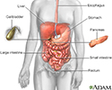
Digestive system - illustration
The esophagus, stomach, large and small intestine, aided by the liver, gallbladder and pancreas convert the nutritive components of food into energy and break down the non-nutritive components into waste to be excreted.
Digestive system
illustration
-
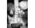
Ileus - X-ray of distended bowel and stomach - illustration
This abdominal X-ray shows a stomach filled with fluid and a swollen (distended) small bowel, caused by a blockage (pseudo-obstruction) in the intestines. A solution containing a dye (barium) that is visible on X-rays was swallowed by the patient (upper GI series).
Ileus - X-ray of distended bowel and stomach
illustration
-
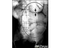
Ileus - X-ray of bowel distension - illustration
This abdominal X-ray shows thickening of the bowel wall and swelling (distention) caused by a blockage (pseudo-obstruction) in the intestines. A solution containing a dye (barium), which is visible on X-ray, was swallowed by the patient (the procedure is known as an upper GI series).
Ileus - X-ray of bowel distension
illustration
-
Intussusception - X-ray - illustration
This abdominal X-ray shows an intestinal condition in which a loop of bowel has slipped into another section of bowel (intussusception), causing swelling, reduced blood flow, obstruction, and tissue damage. Intussusception requires emergency treatment (barium enema or surgery) to prevent intestinal tissue death (necrosis), intestinal perforation, peritonitis, and death.
Intussusception - X-ray
illustration
-
Volvulus - X-ray - illustration
A GI series in a patient with a twisted bowel (volvulus).
Volvulus - X-ray
illustration
-
Small bowel obstruction - X-ray - illustration
X-rays of the abdomen are important in diagnosing the presence of small bowel obstruction. When obstruction occurs, both fluid and gas collect in the intestine. They produce a characteristic pattern called air-fluid levels. The air rises above the fluid and there is a flat surface at the air-fluid interface.
Small bowel obstruction - X-ray
illustration
-
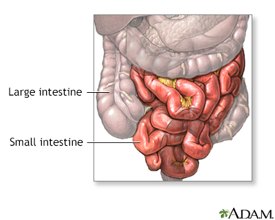
Small bowel resection - series
Presentation
-
Digestive system - illustration
The esophagus, stomach, large and small intestine, aided by the liver, gallbladder and pancreas convert the nutritive components of food into energy and break down the non-nutritive components into waste to be excreted.
Digestive system
illustration
-
Ileus - X-ray of distended bowel and stomach - illustration
This abdominal X-ray shows a stomach filled with fluid and a swollen (distended) small bowel, caused by a blockage (pseudo-obstruction) in the intestines. A solution containing a dye (barium) that is visible on X-rays was swallowed by the patient (upper GI series).
Ileus - X-ray of distended bowel and stomach
illustration
-
Ileus - X-ray of bowel distension - illustration
This abdominal X-ray shows thickening of the bowel wall and swelling (distention) caused by a blockage (pseudo-obstruction) in the intestines. A solution containing a dye (barium), which is visible on X-ray, was swallowed by the patient (the procedure is known as an upper GI series).
Ileus - X-ray of bowel distension
illustration
-
Intussusception - X-ray - illustration
This abdominal X-ray shows an intestinal condition in which a loop of bowel has slipped into another section of bowel (intussusception), causing swelling, reduced blood flow, obstruction, and tissue damage. Intussusception requires emergency treatment (barium enema or surgery) to prevent intestinal tissue death (necrosis), intestinal perforation, peritonitis, and death.
Intussusception - X-ray
illustration
-
Volvulus - X-ray - illustration
A GI series in a patient with a twisted bowel (volvulus).
Volvulus - X-ray
illustration
-
Small bowel obstruction - X-ray - illustration
X-rays of the abdomen are important in diagnosing the presence of small bowel obstruction. When obstruction occurs, both fluid and gas collect in the intestine. They produce a characteristic pattern called air-fluid levels. The air rises above the fluid and there is a flat surface at the air-fluid interface.
Small bowel obstruction - X-ray
illustration
-
Small bowel resection - series
Presentation
Review Date: 5/14/2024
Reviewed By: Jenifer K. Lehrer, MD, Department of Gastroenterology, Aria - Jefferson Health Torresdale, Jefferson Digestive Diseases Network, Philadelphia, PA. Review provided by VeriMed Healthcare Network. Also reviewed by David C. Dugdale, MD, Medical Director, Brenda Conaway, Editorial Director, and the A.D.A.M. Editorial team.


