
Acute cholecystitis
Cholecystitis - acute; Gallstones - acute cholecystitis; Acalculous cholecystitisAcute cholecystitis is sudden swelling and irritation of the gallbladder. It causes severe belly pain.
Causes
The gallbladder is an organ that sits below the liver. It stores bile, which is produced in the liver. Your body uses bile to digest fats in the small intestine.
Acute cholecystitis occurs when bile becomes trapped in the gallbladder. This often happens because a gallstone blocks the cystic duct, the tube through which bile travels into and out of the gallbladder. When a stone blocks this duct, bile builds up, causing irritation and pressure in the gallbladder. This can lead to swelling and infection.
Bile
Bile is a fluid that is made and released by the liver and stored in the gallbladder. Bile helps with digestion. It breaks down fats into fatty acid...

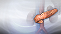
Gallstone
Gallstones are hard deposits that form inside the gallbladder. These may be as small as a grain of sand or as large as a golf ball.
Other causes include:
- Serious illnesses, such as HIV or diabetes
HIV
Human immunodeficiency virus (HIV) is the virus that causes acquired immunodeficiency syndrome (AIDS). When a person becomes infected with HIV, the ...
 ImageRead Article Now Book Mark Article
Diabetes
Diabetes is a long-term (chronic) disease in which the body cannot regulate the amount of sugar in the blood.
 ImageRead Article Now Book Mark Article
 - Tumors of the gallbladder (rare)
Some people are more at risk for gallstones. Risk factors include:
- Being female
- Pregnancy
- Hormone therapy
- Older age
- Being Native American or Hispanic
-
Obesity
Obesity
Obesity means weighing more than what is healthy for a given height. Obesity is a serious, chronic disease. It can lead to other health problems, i...
 ImageRead Article Now Book Mark Article
 - Losing or gaining weight rapidly
- Diabetes
Sometimes, the bile duct becomes blocked temporarily. When this occurs repeatedly, it can lead to long-term (chronic) cholecystitis. This is swelling and irritation that continues over time. Eventually, the gallbladder becomes thick and hard. It does not store and release bile as well as it did.
Long-term (chronic) cholecystitis
Chronic cholecystitis is swelling and irritation of the gallbladder that continues over time. The gallbladder is a sac located under the liver. It s...

Symptoms
The main symptom of acute cholecystitis is pain in the upper right side or upper middle of your belly that usually lasts at least 30 minutes. You may feel:
- Sharp, cramping, or dull pain
- Steady pain
- Pain that spreads to your back or below your right shoulder blade
- Pain that occurs after a meal, more commonly after a fatty meal
Other symptoms that may occur include:
- Clay-colored stools
- Fever
-
Nausea and vomiting
Nausea and vomiting
Nausea is feeling an urge to vomit. It is often called "being sick to your stomach. "Vomiting or throwing-up forces the contents of the stomach up t...
ImageRead Article Now Book Mark Article - Yellowing of the skin and whites of the eyes (jaundice)
Jaundice
Jaundice is a yellow color of the skin, mucus membranes, or eyes. The yellow coloring comes from bilirubin, a byproduct of old red blood cells. Jau...
 ImageRead Article Now Book Mark Article
Exams and Tests
Your health care provider will perform a physical exam and ask about your symptoms. During the physical exam, you will likely have pain when the provider pushes on your belly.
Your provider may order the following blood tests:
-
Amylase and lipase
Amylase
Amylase is an enzyme that helps digest carbohydrates. It is made primarily in the pancreas and the glands that make saliva, and can be found at low ...
 ImageRead Article Now Book Mark Article
 -
Bilirubin
Bilirubin
The bilirubin blood test measures the level of bilirubin in the blood. Bilirubin is a yellowish pigment found in bile, a fluid made by the liver. Bi...
ImageRead Article Now Book Mark Article -
Complete blood count (CBC)
Complete blood count
A complete blood count (CBC) test measures the following:The number of white blood cells (WBC count)The number of red blood cells (RBC count)The numb...
 ImageRead Article Now Book Mark Article
 -
Liver function tests
Liver function tests
Liver function tests are common tests that are used to see how well the liver is working. Tests include:AlbuminAlpha-1 antitrypsinAlkaline phosphata...
 ImageRead Article Now Book Mark Article
Imaging tests can show gallstones or inflammation. You may have one or more of these tests:
-
Abdominal ultrasound
Abdominal ultrasound
Abdominal ultrasound is a type of imaging test. It is used to look at organs in the abdomen, including the liver, gallbladder, pancreas, and kidneys...
 ImageRead Article Now Book Mark Article
 -
Abdominal CT scan or MRI scan
Abdominal CT scan
An abdominal CT scan is an imaging test that uses x-rays to create cross-sectional pictures of the belly area. CT stands for computed tomography....
 ImageRead Article Now Book Mark Article
MRI scan
A magnetic resonance imaging (MRI) scan is an imaging test that uses powerful magnets and radio waves to create pictures of the body. It does not us...
 ImageRead Article Now Book Mark Article
 -
Abdominal x-ray
Abdominal x-ray
An abdominal x-ray is an imaging test to look at organs and structures in the abdomen. Organs include the liver, spleen, stomach, and intestines. T...
 ImageRead Article Now Book Mark Article
 -
Gallbladder radionuclide scan
Gallbladder radionuclide scan
Gallbladder radionuclide scan is a test that uses radioactive material to check gallbladder function. It is also used to look for bile duct blockage...
 ImageRead Article Now Book Mark Article
Treatment
If you have severe belly pain, seek medical attention right away.
In the emergency room, you'll be given fluids through a vein. You may also be given antibiotics to fight infection.
Cholecystitis may clear up on its own. However, if you have gallstones, you will probably need surgery to remove your gallbladder.
Surgery to remove your gallbladder
Open gallbladder removal is surgery to remove the gallbladder through a large cut in your abdomen. The gallbladder is an organ that sits below the li...
Nonsurgical treatment includes:
- Antibiotics you take at home to fight infection
- Low-fat diet (if you are able to eat)
- Pain medicines
You may need emergency surgery if you have complications such as:
- Tissue death (gangrene) of the gallbladder
- A hole that forms in the wall of the gallbladder (perforation)
-
Inflamed pancreas (pancreatitis)
Inflamed pancreas
Acute pancreatitis is sudden swelling and inflammation of the pancreas.
ImageRead Article Now Book Mark Article - Persistent bile duct blockage
- Infection due to a stone in the common bile duct -- also known as cholangitis

If you are very ill, a tube may be placed through your belly into your gallbladder to drain it. Once you feel better, your provider may recommend that you have surgery.
Surgery
Open gallbladder removal is surgery to remove the gallbladder through a large cut in your abdomen.

Outlook (Prognosis)
Most people who have surgery to remove their gallbladder recover completely.
Possible Complications
Untreated, cholecystitis may lead to any of the following health problems:
-
Empyema (pus in the gallbladder)
Empyema
Empyema is a collection of pus in the space between the lung and the inner surface of the chest wall (pleural space).
 ImageRead Article Now Book Mark Article
 - Gangrene
- Injury to the bile ducts draining the liver (may occur after gallbladder surgery)
- Pancreatitis
- Perforation
-
Peritonitis (inflammation of the lining of the abdomen)
Peritonitis
Peritonitis is an inflammation (irritation) of the peritoneum. This is the thin tissue that lines the inner wall of the abdomen and covers most of t...
 ImageRead Article Now Book Mark Article
When to Contact a Medical Professional
Contact your provider if you have:
- Severe belly pain that does not go away
- Symptoms of cholecystitis return
Prevention
Removing the gallbladder and gallstones will prevent further attacks.
References
Clary B, Broderick R, Schaeffer AB, Moffett J, White R. Biliary system. In: Tyler DS, Hayes-Dixon A, Hines J, et al, eds. Sabiston Textbook of Surgery. 22nd ed. Philadelphia, PA: Elsevier; 2026:chap 88.
Fogel EL, Sherman S. Diseases of the gallbladder and bile ducts. In: Goldman L, Cooney KA, eds. Goldman-Cecil Medicine. 27th ed. Philadelphia, PA: Elsevier; 2024:chap 141.
Glasgow RE. Treatment of gallstone disease. In: Feldman M, Friedman LS, Brandt LJ, eds. Sleisenger and Fordtran's Gastrointestinal and Liver Disease. 11th ed. Philadelphia, PA: Elsevier; 2021:chap 66.
Wang DQ-H, Afdhal NH. Gallstone disease. In: Feldman M, Friedman LS, Brandt LJ, eds. Sleisenger and Fordtran's Gastrointestinal and Liver Disease. 11th ed. Philadelphia, PA: Elsevier; 2021:chap 65.
-
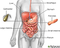
Digestive system - illustration
The esophagus, stomach, large and small intestine, aided by the liver, gallbladder and pancreas convert the nutritive components of food into energy and break down the non-nutritive components into waste to be excreted.
Digestive system
illustration
-
Cholecystitis, CT scan - illustration
This is a CT scan of the upper abdomen showing cholecystitis (gall stones).
Cholecystitis, CT scan
illustration
-
Cholecystitis - cholangiogram - illustration
Cholelithiasis can be seen on a cholangiogram. Radio-opaque dye is used to enhance the x-ray. Multiple stones are present in the gallbladder (PTCA).
Cholecystitis - cholangiogram
illustration
-
Cholecystolithiasis - illustration
Cholecystolithiasis. CT scan of the upper abdomen showing multiple gallstones.
Cholecystolithiasis
illustration
-
Gallstones, cholangiogram - illustration
A cholecystogram in a patient with gallstones.
Gallstones, cholangiogram
illustration
-
Gallbladder removal - Series
Presentation
-
Cholecystogram - illustration
A cholecystogram is an x-ray procedure used to help evaluate the gallbladder. For the procedure, a special diet is consumed prior to the test and contrast tablets are also swallowed to help visualize the gallbladder on x-ray. The test is used to help in diagnosing disorders of the liver and gallbladder, including gallstones and tumors.
Cholecystogram
illustration


 from the liver.</p>")
. Gallstones can affect different locations. Obstruction of the cystic duct leading to severe abdominal pain (biliary colic). Infection or inflammation of the gallbladder (cholecystitis). Blockage of the biliary ducts leading to the duodenum (biliary obstruction). In each case, the gallbladder is often removed (cholecystectomy).</p>")
. Instruments are inserted through 2 more small puncture holes. The gallbladder is found, the vessels and tubes are cut, and the gallbladder is removed.</p>")
 is recommended. A small incision is made just below the rib cage on the right side of the abdomen. The liver is moved to expose the gallbladder. The vessels and tubes (cystic duct and artery) to and from the gallbladder are cut and the gallbladder is removed. The tube (common bile duct) that drains the digestive fluid (bile) from the liver to the small intestine (duodenum) is examined for blockages or stones. A small flat tube may be left in for several days to drain out fluids if there is inflammation or infection.</p>")


-
Digestive system - illustration
The esophagus, stomach, large and small intestine, aided by the liver, gallbladder and pancreas convert the nutritive components of food into energy and break down the non-nutritive components into waste to be excreted.
Digestive system
illustration
-
Cholecystitis, CT scan - illustration
This is a CT scan of the upper abdomen showing cholecystitis (gall stones).
Cholecystitis, CT scan
illustration
-
Cholecystitis - cholangiogram - illustration
Cholelithiasis can be seen on a cholangiogram. Radio-opaque dye is used to enhance the x-ray. Multiple stones are present in the gallbladder (PTCA).
Cholecystitis - cholangiogram
illustration
-
Cholecystolithiasis - illustration
Cholecystolithiasis. CT scan of the upper abdomen showing multiple gallstones.
Cholecystolithiasis
illustration
-
Gallstones, cholangiogram - illustration
A cholecystogram in a patient with gallstones.
Gallstones, cholangiogram
illustration
-
Gallbladder removal - Series
Presentation
-
Cholecystogram - illustration
A cholecystogram is an x-ray procedure used to help evaluate the gallbladder. For the procedure, a special diet is consumed prior to the test and contrast tablets are also swallowed to help visualize the gallbladder on x-ray. The test is used to help in diagnosing disorders of the liver and gallbladder, including gallstones and tumors.
Cholecystogram
illustration
Review Date: 7/22/2025
Reviewed By: Todd Eisner, MD, Private practice specializing in Gastroenterology in Boca Raton and Delray Beach, Florida at Gastroenterology Consultants of Boca Raton. Affiliate Assistant Professor, Florida Atlantic University School of Medicine. Review provided by VeriMed Healthcare Network. Also reviewed by David C. Dugdale, MD, Medical Director, Brenda Conaway, Editorial Director, and the A.D.A.M. Editorial team.
 All rights reserved.