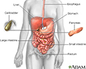
Bleeding esophageal varices
Liver cirrhosis - varices; Cryptogenic chronic liver disease - varices; End-stage liver disease - varices; Alcoholic liver disease - varices; NASH varices; Alcoholic hepatitis - varices; Metabolic dysfunction-associated steatohepatitis (MASH) varicesThe esophagus (food pipe) is the tube that connects your throat to your stomach. Varices are enlarged veins that may be found in the esophagus in people with cirrhosis of the liver. These veins may rupture and bleed.
Causes
Scarring (cirrhosis) of the liver is the most common cause of esophageal varices. This scarring cuts down on blood flowing through the liver. As a result, more blood flows through the veins of the esophagus.
Cirrhosis
Cirrhosis is scarring of the liver and poor liver function. It is the last stage of chronic liver disease.

The extra blood flow causes the veins in the esophagus to balloon outward forming esophageal varices. Heavy bleeding can occur if the varices tear.
Any type of long-term (chronic) liver disease can cause esophageal varices.
Varices can also occur in the upper part of the stomach. These are called gastric varices.
Symptoms
People with chronic liver disease and esophageal varices may have no symptoms.
If there is only a small amount of bleeding, the only symptom may be dark or black streaks in the stools.
If larger amounts of bleeding occur, symptoms may include:
- Black, tarry stools
- Bloody stools
-
Lightheadedness
Lightheadedness
Dizziness is a term that is often used to describe 2 different symptoms: lightheadedness and vertigo. Lightheadedness is a feeling that you might fai...
 ImageRead Article Now Book Mark Article
 - Paleness
- Symptoms of chronic liver disease
-
Vomiting blood
Vomiting blood
Vomiting blood is regurgitating (throwing up) contents of the stomach that contains blood. Vomited blood may appear bright red, dark red, or look lik...
Read Article Now Book Mark Article
Exams and Tests
Your health care provider will do a physical exam which may show:
- Bloody or black stool (in a rectal exam)
-
Low blood pressure
Low blood pressure
Low blood pressure occurs when blood pressure is below normal. This means the heart, brain, and other parts of the body may not get enough blood. I...
 ImageRead Article Now Book Mark Article
 -
Rapid heart rate
Rapid heart rate
A bounding pulse is a strong throbbing felt over one of the arteries in the body. It is due to a forceful heartbeat.
 ImageRead Article Now Book Mark Article
 - Signs of chronic liver disease or cirrhosis
Tests to find the source of the bleeding and check if there is active bleeding include:
-
EGD or upper endoscopy, which involves the use of a camera on a flexible tube to examine the esophagus and stomach.
EGD
Esophagogastroduodenoscopy (EGD) is a test to examine the lining of the esophagus, stomach, and first part of the small intestine (the duodenum)....
 ImageRead Article Now Book Mark Article
 - Insertion of a tube through the nose into the stomach (nasogastric tube) to look for signs of bleeding.
Some providers suggest EGD for people who are newly diagnosed with mild to moderate cirrhosis. This test screens for esophageal varices and treats them before there is bleeding.
Treatment
The goal of treatment is to stop acute bleeding as soon as possible. Bleeding must be controlled quickly to prevent shock and death.
Acute
Acute means sudden. Acute symptoms appear, change, or worsen rapidly. It is the opposite of chronic.

Shock
Shock is a life-threatening condition that occurs when the body is not getting enough blood flow. Lack of blood flow means the cells and organs do n...

If massive bleeding occurs, a person may need to be put on a ventilator to protect their airway and prevent blood from going down into the lungs.
To stop the bleeding, the provider may pass an endoscope (tube with a small light at the end) into the esophagus:
Endoscope
An endoscope is a medical device with a light attached. It is used to look inside a body cavity or organ. The scope is inserted through a natural o...
- A clotting medicine may be injected into the varices.
- A rubber band may be placed around the bleeding varices (called banding or band ligation). Banding is the most common endoscopic treatment for esophageal varices.
Other treatments to stop the bleeding:
- A medicine to tighten blood vessels may be given through a vein. Examples include octreotide or vasopressin.
Tighten blood vessels
Vasoconstriction is the narrowing (constriction) of blood vessels by small muscles in their walls. When blood vessels constrict, blood flow is slowe...
 ImageRead Article Now Book Mark Article
Vasopressin
The antidiuretic blood test measures the level of antidiuretic hormone (ADH) in blood.
Read Article Now Book Mark Article - Rarely, a tube may be inserted through the nose into the stomach and inflated with air. This produces pressure against the bleeding veins (balloon tamponade).
Once the bleeding is stopped, other varices can be treated with medicines and medical procedures to prevent future bleeding. These include:
- Drugs called beta blockers, such as propranolol, nadolol, and carvedilol that reduce portal vein pressure and the risk of bleeding.
- A rubber band can be placed around the varices during an EGD procedure. Also, some medicines can be injected into the varices during EGD to cause them to clot.
-
Transjugular intrahepatic portosystemic shunt (TIPS). This is a procedure to create new connections between two blood vessels in your liver. This can decrease pressure in the varices and prevent bleeding episodes from happening again.
Transjugular intrahepatic portosystemic...
Transjugular intrahepatic portosystemic shunt (TIPS) is a procedure to create new connections between two blood vessels in your liver. You may need ...
 ImageRead Article Now Book Mark Article
In rare cases, emergency surgery may be used to treat people if other treatment fails. Portacaval shunts or surgery to reduce the pressure in the esophageal varices are treatment options, but these procedures are risky.
People with bleeding varices from liver disease may need more treatment for their liver disease, including a liver transplant.
Outlook (Prognosis)
Bleeding often comes back with or without treatment.
Bleeding esophageal varices are a serious complication of liver disease and have a poor outcome.
Placement of a shunt can lead to an increase of metabolic toxins gong to the brain. This can lead to mental status changes.
Possible Complications
Future problems caused by varices may include:
- Narrowing or stricture of the esophagus due to scarring after a procedure
Stricture of the esophagus
Benign esophageal stricture is a narrowing of the esophagus (the tube from the mouth to the stomach). It causes swallowing difficulties. Benign mean...
Read Article Now Book Mark Article - Return of bleeding after treatment
When to Contact a Medical Professional
Contact your provider or go to an emergency room if you vomit blood or have black tarry stools.
Prevention
Treating the causes of liver disease may prevent bleeding. Liver transplantation should be considered for some people.
References
Garcia-Tsao G. Cirrhosis and its sequelae. In: Goldman L, Cooney KA, eds. Goldman-Cecil Medicine. 27th ed. Philadelphia, PA: Elsevier; 2024:chap 139.
Savides TJ, Jensen DM. Gastrointestinal bleeding. In: Feldman M, Friedman LS, Brandt LJ, eds. Sleisenger and Fordtran's Gastrointestinal and Liver Disease: Pathophysiology/Diagnosis/Management. 11th ed. Philadelphia, PA: Elsevier; 2021:chap 20.
Shah VH, Kamath PS. Portal hypertension and variceal bleeding. In: Feldman M, Friedman LS, Brandt LJ, eds. Sleisenger and Fordtran's Gastrointestinal and Liver Disease: Pathophysiology/Diagnosis/Management. 11th ed. Philadelphia, PA: Elsevier; 2021:chap 92.
-
Digestive system - illustration
The esophagus, stomach, large and small intestine, aided by the liver, gallbladder and pancreas convert the nutritive components of food into energy and break down the non-nutritive components into waste to be excreted.
Digestive system
illustration
-
Liver blood supply - illustration
The proper hepatic artery supplies blood to the liver.
Liver blood supply
illustration
-
Digestive system - illustration
The esophagus, stomach, large and small intestine, aided by the liver, gallbladder and pancreas convert the nutritive components of food into energy and break down the non-nutritive components into waste to be excreted.
Digestive system
illustration
-
Liver blood supply - illustration
The proper hepatic artery supplies blood to the liver.
Liver blood supply
illustration
Review Date: 12/31/2023
Reviewed By: Jenifer K. Lehrer, MD, Department of Gastroenterology, Aria - Jefferson Health Torresdale, Jefferson Digestive Diseases Network, Philadelphia, PA. Review provided by VeriMed Healthcare Network. Also reviewed by David C. Dugdale, MD, Medical Director, Brenda Conaway, Editorial Director, and the A.D.A.M. Editorial team.


