Renal papillary necrosis
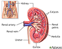
Necrosis - renal papillae; Renal medullary necrosisRenal papillary necrosis is a disorder of the kidneys in which all or part of the renal papillae die. The renal papillae are the areas where the openings of the collecting ducts enter the kidney and where urine flows into the ureters.
Causes
Renal papillary necrosis often occurs with analgesic nephropathy. This is damage to one or both kidneys caused by overexposure to pain medicines. But, other conditions can also cause renal papillary necrosis, including:
Analgesic nephropathy
Analgesic nephropathy involves damage to one or both kidneys caused by overexposure to mixtures of medicines, especially over-the-counter pain medici...

-
Diabetic nephropathy
Diabetic nephropathy
Kidney disease or kidney damage often occurs over time in people with diabetes. This type of kidney disease is called diabetic nephropathy.
 ImageRead Article Now Book Mark Article
 - Kidney infection (pyelonephritis)
- Kidney transplant rejection
-
Sickle cell anemia, a common cause of renal papillary necrosis in children
Sickle cell anemia
Sickle cell disease is a disorder passed down through families. The red blood cells that are normally shaped like a disk take on a sickle or crescen...
 ImageRead Article Now Book Mark Article
 - Urinary tract blockage
Symptoms
Symptoms of renal papillary necrosis may include:
- Back pain or flank pain
Flank pain
Flank pain is pain in one side of the body between the upper belly area (abdomen) and the back.
 ImageRead Article Now Book Mark Article
 -
Bloody, cloudy, or dark urine
Bloody, cloudy, or dark urine
Blood in your urine is called hematuria. The amount may be very small and only detected with urine tests or under a microscope. In other cases, the...
 ImageRead Article Now Book Mark Article
 - Tissue pieces in the urine
Other symptoms that may occur with this disease:
- Fever and chills
-
Painful urination
Painful urination
Painful urination is any pain, discomfort, or burning sensation when passing urine.
 ImageRead Article Now Book Mark Article
 - Needing to urinate more often than usual (frequent urination) or a sudden, strong urge to urinate (urgency)
- Difficulty starting or maintaining a urine stream (urinary hesitancy)
Urinary hesitancy
Difficulty starting or maintaining a urine stream is called urinary hesitancy.
 ImageRead Article Now Book Mark Article
 -
Urinary incontinence
Urinary incontinence
Urinary (or bladder) incontinence occurs when you are not able to keep urine from leaking out of your urethra. The urethra is the tube that carries ...
 ImageRead Article Now Book Mark Article
 - Urinating large amounts
- Urinating often at night
Exams and Tests
The area over the affected kidney (in the flank) may feel tender during an exam. There may be a history of urinary tract infections. There may be signs of blocked urine flow or kidney failure.
Blocked urine flow
Obstructive uropathy is a condition in which the flow of urine is blocked. This causes the urine to back up and injure one or both kidneys.

Tests that may be done include:
- Urine test
- Blood tests
- Ultrasound, CT, or other imaging tests of the kidneys
Treatment
There is no specific treatment for renal papillary necrosis. Treatment depends on the cause. For example, if analgesic nephropathy is the cause, your health care provider will recommend that you stop using the medicine that is causing it. This may allow the kidney to heal over time.
Outlook (Prognosis)
How well a person does, depends on what is causing the condition. If the cause can be controlled, the condition may go away on its own. Sometimes, people with this condition develop kidney failure and will need dialysis or a kidney transplant.
Dialysis
Dialysis treats end-stage kidney disease also called kidney failure. It removes waste from your blood when your kidneys can no longer do their job. ...

Kidney transplant
A kidney transplant is surgery to place a healthy kidney into a person with kidney failure.

Possible Complications
Health problems that may result from renal papillary necrosis include:
- Kidney infection
- Kidney stones
- Kidney cancer, especially in people who take a lot of pain medicines
When to Contact a Medical Professional
Contact your provider for an appointment if:
- You have bloody urine
Bloody urine
Blood in your urine is called hematuria. The amount may be very small and only detected with urine tests or under a microscope. In other cases, the...
 ImageRead Article Now Book Mark Article
 - You develop other symptoms of renal papillary necrosis, especially after taking over-the-counter pain medicines
Prevention
Controlling diabetes or sickle cell anemia may reduce your risk. To prevent renal papillary necrosis from analgesic nephropathy, follow your provider's instructions when using medicines, including over-the-counter pain relievers. Do not take more than the recommended dose without asking your provider.
References
Chen W, Bushinsky DA. Nephrolithiasis and nephrocalcinosis. In: Feehally J, Floege J, Tonelli M, Johnson RJ, eds. Comprehensive Clinical Nephrology. 7th ed. Philadelphia, PA: Elsevier; 2024:chap 60.
Cooper KL, Badalato GM, Rutman MP. Infections of the urinary tract. In: Partin AW, Dmochowski RR, Kavoussi LR, Peters CA, eds. Campbell-Walsh-Wein Urology. 12th ed. Philadelphia, PA: Elsevier; 2021:chap 55.
Gharavi AG,Landry DW. Approach to the patient with renal disease. In: Goldman L, Cooney KA, eds. Goldman-Cecil Medicine. 27th ed. Philadelphia, PA: Elsevier; 2024:chap 100.
-
Kidney anatomy - illustration
The kidneys are responsible for removing wastes from the body, regulating electrolyte balance and blood pressure, and the stimulation of red blood cell production.
Kidney anatomy
illustration
-
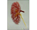
Kidney - blood and urine flow - illustration
This is the typical appearance of the blood vessels (vasculature) and urine flow pattern in the kidney. The blood vessels are shown in red and the urine flow pattern in yellow.
Kidney - blood and urine flow
illustration
-
Kidney anatomy - illustration
The kidneys are responsible for removing wastes from the body, regulating electrolyte balance and blood pressure, and the stimulation of red blood cell production.
Kidney anatomy
illustration
-
Kidney - blood and urine flow - illustration
This is the typical appearance of the blood vessels (vasculature) and urine flow pattern in the kidney. The blood vessels are shown in red and the urine flow pattern in yellow.
Kidney - blood and urine flow
illustration
Review Date: 8/28/2023
Reviewed By: Walead Latif, MD, Nephrologist and Clinical Associate Professor, Rutgers Medical School, Newark, NJ. Review provided by VeriMed Healthcare Network. Also reviewed by David C. Dugdale, MD, Medical Director, Brenda Conaway, Editorial Director, and the A.D.A.M. Editorial team.


