Acute myeloid leukemia - adult
Acute myelogenous leukemia; AML; Acute granulocytic leukemia; Acute nonlymphocytic leukemia (ANLL); Leukemia - acute myeloid (AML); Leukemia - acute granulocytic; Leukemia - nonlymphocytic (ANLL)Acute myeloid leukemia (AML) is cancer that starts inside bone marrow. This is the soft tissue in the center of bones that helps form all blood cells. The cancer grows from cells that would normally turn into white blood cells.
Acute means the disease grows quickly and usually has an aggressive course.
Causes
AML is one of the most common types of leukemia among adults.
Leukemia
Leukemia is a type of blood cancer that begins in the bone marrow. Bone marrow is the soft tissue in the center of the bones, where blood cells are ...

AML is more common in men than women.
The bone marrow helps the body fight infections and makes other blood components. People with AML have many abnormal immature white blood cells inside their bone marrow. The cells grow very quickly, and replace healthy blood cells. As a result, people with AML are more likely to have infections. They also have an increased risk of bleeding as the numbers of healthy blood cells decrease.
Most of the time, a health care provider cannot tell you what caused AML. However, the following things can lead to some types of leukemia, including AML:
-
Blood disorders, including polycythemia vera, essential thrombocythemia, and myelodysplasia
Polycythemia vera
Polycythemia vera (PV) is a bone marrow disease that leads to an abnormal increase in the number of blood cells. The red blood cells are the most af...
Read Article Now Book Mark ArticleEssential thrombocythemia
Essential thrombocythemia (ET) is a condition in which the bone marrow produces too many platelets. Platelets are particles in the blood that aid in...
 ImageRead Article Now Book Mark Article
 - Certain chemicals (for example, benzene)
-
Certain chemotherapy drugs, including etoposide and drugs known as alkylating agents
Chemotherapy
The term chemotherapy is used to describe cancer-killing drugs. Chemotherapy may be used to:Cure the cancerShrink the cancerPrevent the cancer from ...
 ImageRead Article Now Book Mark Article
 - Exposure to certain chemicals and harmful substances
- Radiation
- Weak immune system due to an organ transplant
Problems with your genes may also cause AML to develop.
Symptoms
Symptoms of AML are mainly due to the effects on blood elements. Symptoms of AML may include any of the following:
-
Bleeding from the nose
Bleeding from the nose
A nosebleed is loss of blood from the tissue lining the nose. Bleeding most often occurs from one nostril only.
 ImageRead Article Now Book Mark Article
 -
Bleeding and swelling (rare) in the gums
Bleeding
Bleeding gums can be a sign that you have or may develop gum disease. Ongoing gum bleeding may be due to plaque buildup on the teeth. It can also b...
Read Article Now Book Mark ArticleSwelling
Swollen gums are abnormally enlarged, bulging, or protruding gums.
 ImageRead Article Now Book Mark Article
 - Bruising
-
Bone pain or tenderness
Bone pain or tenderness
Bone pain or tenderness is aching or other discomfort in one or more bones.
 ImageRead Article Now Book Mark Article
 - Fever and fatigue
- Heavy menstrual periods
- Pale skin
-
Shortness of breath (gets worse with exercise)
Shortness of breath
Breathing difficulty may involve:Difficult breathing Uncomfortable breathingFeeling like you are not getting enough air
 ImageRead Article Now Book Mark Article
 - Weight loss
Exams and Tests
The provider will perform a physical exam. There may be signs of a swollen spleen, liver, or lymph nodes. Tests done include:
- A complete blood count (CBC) may show anemia and a low number of platelets. A white blood cell count (WBC) can be high, low, or normal.
CBC
A complete blood count (CBC) test measures the following:The number of white blood cells (WBC count)The number of red blood cells (RBC count)The numb...
 ImageRead Article Now Book Mark Article
WBC
A WBC count is a blood test to measure the number of white blood cells (WBCs) in the blood. It is a part of a complete blood count (CBC). WBCs are a...
 ImageRead Article Now Book Mark Article
 -
Bone marrow aspiration and biopsy will show if there are any leukemia cells.
Bone marrow aspiration
Bone marrow is the soft tissue inside bones that helps form blood cells. It is found in the hollow part of most bones. Bone marrow aspiration is th...
 ImageRead Article Now Book Mark Article
Biopsy
A bone marrow biopsy is the removal of marrow from inside one of your bones. Bone marrow is the soft tissue inside bones that helps form blood cells...
 ImageRead Article Now Book Mark Article
If your provider learns you do have this type of leukemia, further tests will be done to determine the specific type of AML. Subtypes are based on specific changes in genes (mutations) and how the leukemia cells appear under the microscope.
Treatment
Treatment involves using medicines (chemotherapy) to kill the cancer cells. Most types of AML are treated with more than one chemotherapy medicine. Drugs that are targeted to specific mutations in the leukemic cells are also often used.
Chemotherapy
The term chemotherapy is used to describe cancer-killing drugs. Chemotherapy may be used to:Cure the cancerShrink the cancerPrevent the cancer from ...

Chemotherapy kills normal cells, too. This may cause side effects such as:
- Increased risk of bleeding
Risk of bleeding
Your bone marrow makes cells called platelets. These cells keep you from bleeding too much by helping your blood clot. Chemotherapy, radiation, and...
Read Article Now Book Mark Article - Increased risk for infection (your doctor may want you to keep away from other people to prevent infection)
- Weight loss (you will need to eat extra calories)
You will need to eat extra calories
If you are sick or undergoing cancer treatment, you may not feel like eating. But it is important to get enough protein and calories so you do not l...
Read Article Now Book Mark Article -
Mouth sores
Mouth sores
Oral mucositis is tissue swelling and irritation in the mouth. Radiation therapy or chemotherapy may cause mucositis. Follow your health care provi...
Read Article Now Book Mark Article
Other supportive treatments for AML may include:
- Antibiotics to treat infection
- Red blood cell transfusions to fight anemia
Anemia
Anemia is a condition in which the body does not have enough healthy red blood cells. Red blood cells provide oxygen to body tissues. Different type...
 ImageRead Article Now Book Mark Article
 -
Platelet transfusions to control bleeding
Platelet
A platelet count is a lab test to measure how many platelets you have in your blood. Platelets are particles in the blood that help the blood clot. ...
Read Article Now Book Mark Article
A bone marrow (stem cell) transplant may be tried. This decision is decided by several factors, including:
Bone marrow (stem cell) transplant
A bone marrow transplant is a procedure to replace damaged or diseased bone marrow with healthy bone marrow stem cells. Bone marrow is the soft, fatt...

- Your age and overall health
- Certain genetic changes in the leukemia cells
- The availability of donors
Support Groups
You can ease the stress of illness by joining a cancer support group. Sharing with others who have common experiences and problems can help you not feel alone.
Support group
The following organizations are good resources for information on cancer:American Cancer Society. Support and online communities. www. cancer. org/...
Outlook (Prognosis)
When a bone marrow biopsy shows no evidence of AML, you are said to be in remission. How well you do depends on your overall health and the genetic subtype of the AML cells.
Remission is not the same as a cure. More therapy is usually needed, either in the form of more chemotherapy or a bone marrow transplant.
With treatment, younger people with AML tend to do better than those who develop the disease at an older age. The 5-year survival rate is much lower in older adults than in younger people. Experts say this is partly due to the fact that younger people are better able to tolerate strong chemotherapy medicines. Also, leukemia in older people tends to be more resistant to current treatments.
If the cancer does not come back (relapse) within 5 years of the diagnosis, you are likely cured.
When to Contact a Medical Professional
Contact your provider to schedule an appointment if you:
- Develop symptoms of AML
- Have AML and have a fever that will not go away or other signs of infection
Prevention
If you work around radiation or chemicals linked to leukemia, always wear protective gear.
References
Appelbaum FR. Acute leukemias in adults. In: Niederhuber JE, Armitage JO, Kastan MB, Doroshow JH, Tepper JE, eds. Abeloff's Clinical Oncology. 6th ed. Philadelphia, PA: Elsevier; 2020:chap 95.
Erba HP. Clinical manifestations and treatment of acute myeloid leukemia. In: Hoffman R, Benz EJ, Silberstein LE, et al, eds. Hematology: Basic Principles and Practice. 8th ed. Philadelphia, PA: Elsevier; 2023:chap 60.
National Cancer Institute website. Adult acute myeloid leukemia treatment (PDQ) -- health professional version. www.cancer.gov/types/leukemia/hp/adult-aml-treatment-pdq. Updated March 6, 2024. Accessed June 18, 2024.
-
Auer rods - illustration
Note multiple Auer rods which are found only in acute myeloid leukemias, either myeloblastic or monoblastic. These rods consist of clumps of azurophilic granule material.
Auer rods
illustration
-
Acute monocytic leukemia - skin - illustration
Acute monocytic leukemia. These lesions are rarely found in chronic leukemia but are a common finding in acute forms. They appear as erythematous infiltrations of the skin, forming papules, macules, and plaques. Pruritus may be present.
Acute monocytic leukemia - skin
illustration
-
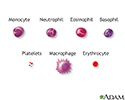
Blood cells - illustration
Blood is comprised of red blood cells, platelets, and various white blood cells.
Blood cells
illustration
-
Auer rods - illustration
Note multiple Auer rods which are found only in acute myeloid leukemias, either myeloblastic or monoblastic. These rods consist of clumps of azurophilic granule material.
Auer rods
illustration
-
Acute monocytic leukemia - skin - illustration
Acute monocytic leukemia. These lesions are rarely found in chronic leukemia but are a common finding in acute forms. They appear as erythematous infiltrations of the skin, forming papules, macules, and plaques. Pruritus may be present.
Acute monocytic leukemia - skin
illustration
-
Blood cells - illustration
Blood is comprised of red blood cells, platelets, and various white blood cells.
Blood cells
illustration
Review Date: 6/17/2024
Reviewed By: Todd Gersten, MD, Hematology/Oncology, Florida Cancer Specialists & Research Institute, Wellington, FL. Review provided by VeriMed Healthcare Network. Also reviewed by David C. Dugdale, MD, Medical Director, Brenda Conaway, Editorial Director, and the A.D.A.M. Editorial team.


