Vaginal dryness
Vaginitis - atrophic; Vaginitis due to reduced estrogen; Atrophic vaginitis; Menopause vaginal drynessVaginal dryness is present when the tissues of the vagina are not well-lubricated and healthy.
Causes
Atrophic vaginitis is caused by a decrease in estrogen.
Estrogen keeps the tissues of the vagina lubricated and healthy. Normally, the lining of the vagina makes a clear, lubricating fluid. This fluid makes sexual intercourse more comfortable. It also helps decrease vaginal dryness.
If estrogen levels drop off, the tissues of the vagina shrink and become thinner. This causes dryness and inflammation.
Estrogen levels normally drop after menopause. The following may also cause estrogen levels to drop:
- Medicines or hormones used in the treatment of breast cancer, endometriosis, fibroids, or infertility
- Surgery to remove the ovaries
- Radiation treatment to the pelvic area
- Chemotherapy
- Severe stress, depression
- Smoking
Some women develop this problem right after childbirth or while breastfeeding. Estrogen levels are lower at these times.
The vagina can also become further irritated from soaps, laundry detergents, lotions, perfumes, or douches. Certain medicines, smoking, tampons, and condoms may also cause or worsen vaginal dryness.
Vagina can also become further irritate
Vulvovaginitis or vaginitis is swelling or infection of the vulva and vagina. Vaginitis is a common problem that can affect women and girls of all ag...
Symptoms
Symptoms include:
- Burning on urination
- Light bleeding after intercourse
Bleeding
Bleeding is the loss of blood. Bleeding may be:Inside the body (internal)Outside the body (external)Bleeding may occur:Inside the body when blood le...
 ImageRead Article Now Book Mark Article
 - Painful sexual intercourse
- Slight vaginal discharge
Vaginal discharge
Vaginal discharge refers to secretions from the vagina. The discharge may be:Thick, pasty, or thinClear, cloudy, bloody, white, yellow, or greenOdor...
 ImageRead Article Now Book Mark Article
 - Vaginal soreness, itching or burning
Itching
Itching is a tingling or irritation of the skin that makes you want to scratch the area. Itching may occur all over the body or only in one location...
 ImageRead Article Now Book Mark Article
Exams and Tests
A pelvic exam shows that the walls of the vagina are thin, pale or red.
Your vaginal discharge may be tested to check for other causes for the condition. You may also have hormone level tests to find out if you are in menopause.
Treatment
There are many treatments for vaginal dryness. Before treating your symptoms on your own, a health care provider must find out the cause of the problem.
- Try using lubricants and vaginal moisturizing creams. They will often moisten the area for several hours, up to a day. These can be bought without prescription.
Vaginal moisturizing creams
Question: Is there a drug-free treatment for vaginal dryness? Answer: There are many causes of vaginal dryness. It may be caused by reduced estrogen...
Read Article Now Book Mark Article - Use of a water-soluble vaginal lubricant during intercourse may help. Products with petroleum jelly, mineral oil, or other oils may damage latex condoms or diaphragms.
- Avoid scented soaps, lotions, perfumes, or douches.
Prescription estrogen can work well to treat atrophic vaginitis. It is available as a cream, tablet, suppository, or ring. All of these are placed directly into the vagina. These medicines deliver estrogen directly to the vaginal area. Only a little estrogen is absorbed into the bloodstream. The use of topical vaginal estrogen may also reduce your chances of developing a urinary tract infection. That is particularly true if you have a history of recurrent urinary tract infections.
You may take estrogen (hormone therapy) in the form of a skin patch, or in a pill that you take by mouth if you have hot flashes or other symptoms of menopause. The pill or patch may not provide adequate estrogen to treat your vaginal dryness. In such cases, you may need to add a vaginal hormone medicine as well. If so, talk to your provider about this.
Hormone therapy
Hormone therapy (HT) uses one or more hormones to treat symptoms of menopause. HT uses estrogen, progestin (a type of progesterone), or both. Somet...
You should discuss the risks and benefits of estrogen replacement therapy with your provider.
Outlook (Prognosis)
Proper treatment will ease symptoms most of the time.
Possible Complications
Vaginal dryness can:
- Make you more likely to get yeast or bacterial infections of the vagina.
- Cause sores or cracks in the walls of the vagina.
- Cause pain with sexual intercourse, which may affect your relationship with your partner or spouse. (Talking openly with your partner may help.)
- Increase your risk of developing urinary tract infections (UTI).
When to Contact a Medical Professional
Contact your provider if you have vaginal dryness or soreness, burning, itching, or painful sexual intercourse that does not go away when you use a water-soluble lubricant.
References
Ball JW, Dains JE, Flynn JA, Solomon BS, Stewart RW. Female genitalia. In: Ball JW, Dains JE, Flynn JA, Solomon BS, Stewart RW, eds. Seidel's Guide to Physical Examination. 10th ed. St Louis, MO: Elsevier; 2023:chap 19.
Eckert LO, Lentz GM. Genital tract infections: vulva, vagina, cervix, toxic shock syndrome, endometritis, and salpingitis. In: Gershenson DM, Lentz GM, Valea FA, Lobo RA, eds. Comprehensive Gynecology. 8th ed. Philadelphia, PA: Elsevier; 2022:chap 23.
Lobo RA. Menopause and care of the mature woman: endocrinology, consequences of estrogen deficiency, effects of hormone therapy, and other treatment options. In: Gershenson DM, Lentz GM, Valea FA, Lobo RA, eds. Comprehensive Gynecology. 8th ed. Philadelphia, PA: Elsevier; 2022:chap 14.
Santoro N, Neal-Perry G. Menopause. In: Goldman L, Cooney KA, eds. Goldman-Cecil Medicine. 27th ed. Philadelphia, PA: Elsevier; 2024:chap 222.
-
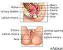
Female reproductive anatomy - illustration
Internal structures of the female reproductive anatomy include the uterus, ovaries, and cervix. External structures include the labium minora and majora, the vagina and the clitoris.
Female reproductive anatomy
illustration
-
Causes of painful intercourse - illustration
Dyspareunia (painful intercourse) refers to pain in the pelvic area during or after intercourse, and can occur in both women and men. Besides possible physical causes, pain may occur in association with psychological factors such as previous sexual trauma.
Causes of painful intercourse
illustration
-
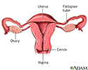
Uterus - illustration
The uterus is a hollow muscular organ located in the female pelvis between the bladder and rectum. The ovaries produce the eggs that travel through the fallopian tubes. Once the egg has left the ovary it can be fertilized and implant itself in the lining of the uterus. The main function of the uterus is to nourish the developing fetus prior to birth.
Uterus
illustration
-
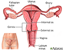
Normal uterine anatomy (cut section) - illustration
The uterus is a muscular organ with thick walls, two upper openings to the fallopian tubes and an inferior opening to the vagina.
Normal uterine anatomy (cut section)
illustration
-
Vaginal atrophy - illustration
Vaginal atrophy is caused by a drop in estrogen levels. This most often occurs during menopause. In normal conditions, the vaginal wall is elastic and thick, and its lining is ridged and lubricated. In vaginal atrophy, the vagina wall shrinks, and its lining becomes pale, smooth, and dry.
Vaginal atrophy
illustration
-
Female reproductive anatomy - illustration
Internal structures of the female reproductive anatomy include the uterus, ovaries, and cervix. External structures include the labium minora and majora, the vagina and the clitoris.
Female reproductive anatomy
illustration
-
Causes of painful intercourse - illustration
Dyspareunia (painful intercourse) refers to pain in the pelvic area during or after intercourse, and can occur in both women and men. Besides possible physical causes, pain may occur in association with psychological factors such as previous sexual trauma.
Causes of painful intercourse
illustration
-
Uterus - illustration
The uterus is a hollow muscular organ located in the female pelvis between the bladder and rectum. The ovaries produce the eggs that travel through the fallopian tubes. Once the egg has left the ovary it can be fertilized and implant itself in the lining of the uterus. The main function of the uterus is to nourish the developing fetus prior to birth.
Uterus
illustration
-
Normal uterine anatomy (cut section) - illustration
The uterus is a muscular organ with thick walls, two upper openings to the fallopian tubes and an inferior opening to the vagina.
Normal uterine anatomy (cut section)
illustration
-
Vaginal atrophy - illustration
Vaginal atrophy is caused by a drop in estrogen levels. This most often occurs during menopause. In normal conditions, the vaginal wall is elastic and thick, and its lining is ridged and lubricated. In vaginal atrophy, the vagina wall shrinks, and its lining becomes pale, smooth, and dry.
Vaginal atrophy
illustration
Review Date: 7/12/2023
Reviewed By: John D. Jacobson, MD, Professor Emeritus, Department of Obstetrics and Gynecology, Loma Linda University School of Medicine, Loma Linda, CA. Also reviewed by David C. Dugdale, MD, Medical Director, Brenda Conaway, Editorial Director, and the A.D.A.M. Editorial team.


