Ectopic pregnancy
Tubal pregnancy; Cervical pregnancy; Tubal ligation - ectopic pregnancyAn ectopic pregnancy is a pregnancy that occurs outside the womb (uterus).
Causes
In most pregnancies, the fertilized egg travels through the fallopian tube to the womb (uterus). If the movement of the egg is blocked or slowed through the tubes, it can lead to an ectopic pregnancy. Things that may cause this problem include:
- Birth defect in the fallopian tubes
- Scarring after a ruptured appendix
Ruptured appendix
Appendicitis is a condition in which your appendix gets inflamed. The appendix is a small pouch attached to the end of the large intestine.
 ImageRead Article Now Book Mark Article
 -
Endometriosis
Endometriosis
Endometriosis occurs when cells from the lining of your womb (uterus) grow in other areas of your body. This can cause pain, heavy vaginal bleeding,...
 ImageRead Article Now Book Mark Article
 - Having had an ectopic pregnancy in the past
- Scarring from past infections or surgery of the female organs
- History of prior abdominal surgery
The following also increase risk for an ectopic pregnancy:
- Age over 35
- Getting pregnant while having an intrauterine device (IUD)
- Having your fallopian tubes tied
- Having had surgery to untie fallopian tubes to become pregnant
- Having had many sexual partners
- Sexually transmitted infections (STI)
- Some infertility treatments
Sometimes, the cause is not known. Hormones may play a role.
The most common site for an ectopic pregnancy is the fallopian tube. In rare cases, this can occur in the ovary, abdomen, or cervix.
Cervix
The cervix is the lower end of the womb (uterus). It is at the top of the vagina. It is about 2. 5 to 3. 5 centimeters (1 to 1. 3 inches) long. Th...

An ectopic pregnancy can occur even if you use birth control.
Symptoms
Symptoms of ectopic pregnancy may include:
- Abnormal vaginal bleeding
- Mild cramping on one side of the pelvis
- No periods
- Pain in the lower belly or pelvic area
If the area around the abnormal pregnancy ruptures and bleeds, symptoms may get worse. They may include:
- Fainting or feeling faint
- Intense pressure in the rectum
- Low blood pressure
- Pain in the shoulder area
- Severe, sharp, and sudden pain in the lower abdomen
Exams and Tests
The health care provider will do a pelvic exam. The exam may show tenderness in the pelvic area.
A pregnancy test and vaginal ultrasound will be done.
Human chorionic gonadotropin (hCG) is a hormone that is produced during pregnancy. Checking the blood level of this hormone can detect pregnancy.
- When hCG levels are above a certain value, a pregnancy sac in the uterus should be seen with ultrasound.
- If the sac is not seen, this may indicate that an ectopic pregnancy is present.
You may need more than one exam, ultrasound, and blood test. Your provider will instruct you about signs to watch for until your next visit.
Treatment
Ectopic pregnancy may be life threatening. The pregnancy cannot continue to birth (term). Effective treatment requires either medical treatment to end the pregnancy or surgical removal of the pregnancy.
If the ectopic pregnancy has not ruptured, treatment may include:
-
Surgery
Surgery
Pelvic laparoscopy is surgery to examine the pelvic organs. It uses a viewing tool called a laparoscope. The surgery is also used to treat certain ...
 ImageRead Article Now Book Mark Article
 - Medicine that ends the pregnancy, along with close monitoring by your provider
You will need emergency medical help if the area of the ectopic pregnancy breaks open (ruptures). Rupture can lead to bleeding and shock. Treatment for shock may include:
- Blood transfusion
- Fluids given through a vein
- Keeping warm
- Oxygen
- Raising the legs
If there is a rupture, surgery is done to stop blood loss and remove the pregnancy. In some cases, the surgeon may have to remove the fallopian tube.
Outlook (Prognosis)
If diagnosed early, treatment is very effective. It's important to seek early care whenever you believe you may be pregnant so your provider may determine the location of the pregnancy.
One out of three women who have had one ectopic pregnancy can have a baby in the future. Another ectopic pregnancy is more likely to occur. Some women do not become pregnant again.
The likelihood of a successful pregnancy after an ectopic pregnancy depends on:
- The woman's age
- Whether she has already had children
- Why the first ectopic pregnancy occurred
- The health of her fallopian tubes
When to Contact a Medical Professional
Contact your provider if you have:
- Abnormal vaginal bleeding
- Lower abdominal or pelvic pain or
- Suspect you might be pregnant
Prevention
Most forms of ectopic pregnancy that occur outside the fallopian tubes are probably not preventable. You may be able to reduce your risk by avoiding conditions that may scar the fallopian tubes. These steps include:
- Practicing safer sex by taking steps before and during sex, which can prevent you from getting an infection
- Getting early diagnosis and treatment of all STIs
- Stopping smoking
References
Alur-Gupta S, Cooney LG, Senapati S, Sammel MD, Barnhart KT. Two-dose versus single-dose methotrexate for treatment of ectopic pregnancy: a meta-analysis. Am J Obstet Gynecol. 2019;221(2):95-108.e2. PMID: 30629908 pubmed.ncbi.nlm.nih.gov/30629908/.
Henn MC, Lall MD. Complications of pregnancy. In: Walls RM, eds. Rosen's Emergency Medicine: Concepts and Clinical Practice. 8th ed. Philadelphia, PA: Elsevier; 2023:chap 173.
Hur HC, Lobo RA. Ectopic pregnancy: etiology, pathology, diagnosis, management, fertility prognosis. In: Gershenson DM, Lentz GM, Valea FA, Lobo RA, eds. Comprehensive Gynecology. 8th ed. Philadelphia, PA: Elsevier; 2022:chap 17.
Nelson AL, Gambone JC. Ectopic pregnancy. In: Hacker NF, Gambone JC, Hobel CJ, eds. Hacker & Moore's Essentials of Obstetrics and Gynecology. 6th ed. Philadelphia, PA: Elsevier; 2016:chap 24.
-
Pelvic laparoscopy - illustration
Laparoscopy is performed when less-invasive surgery is desired. It is also called Band-Aid surgery because only small incisions need to be made to accommodate the small surgical instruments that are used to view the abdominal contents and perform the surgery.
Pelvic laparoscopy
illustration
-
Ultrasound in pregnancy - illustration
The ultrasound has become a standard procedure used during pregnancy. It can demonstrate fetal growth and can detect increasing numbers of conditions including meningomyelocele, congenital heart disease, kidney abnormalities, hydrocephalus, anencephaly, club feet, and other deformities. Ultrasound does not produce ionizing radiation and is considered a very safe procedure for both the mother and the fetus.
Ultrasound in pregnancy
illustration
-
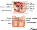
Female reproductive anatomy - illustration
Internal structures of the female reproductive anatomy include the uterus, ovaries, and cervix. External structures include the labium minora and majora, the vagina and the clitoris.
Female reproductive anatomy
illustration
-
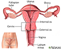
Uterus - illustration
The uterus is a hollow muscular organ located in the female pelvis between the bladder and rectum. The ovaries produce the eggs that travel through the fallopian tubes. Once the egg has left the ovary it can be fertilized and implant itself in the lining of the uterus. The main function of the uterus is to nourish the developing fetus prior to birth.
Uterus
illustration
-
Ectopic pregnancy - illustration
An ectopic pregnancy is one in which the fertilized egg implants in tissue outside of the uterus and the placenta and fetus begin to develop there. The most common site is within a Fallopian tube, however, ectopic pregnancies can occur in the ovary, the abdomen, and in the lower portion of the uterus (the cervix).
Ectopic pregnancy
illustration
-
Pelvic laparoscopy - illustration
Laparoscopy is performed when less-invasive surgery is desired. It is also called Band-Aid surgery because only small incisions need to be made to accommodate the small surgical instruments that are used to view the abdominal contents and perform the surgery.
Pelvic laparoscopy
illustration
-
Ultrasound in pregnancy - illustration
The ultrasound has become a standard procedure used during pregnancy. It can demonstrate fetal growth and can detect increasing numbers of conditions including meningomyelocele, congenital heart disease, kidney abnormalities, hydrocephalus, anencephaly, club feet, and other deformities. Ultrasound does not produce ionizing radiation and is considered a very safe procedure for both the mother and the fetus.
Ultrasound in pregnancy
illustration
-
Female reproductive anatomy - illustration
Internal structures of the female reproductive anatomy include the uterus, ovaries, and cervix. External structures include the labium minora and majora, the vagina and the clitoris.
Female reproductive anatomy
illustration
-
Uterus - illustration
The uterus is a hollow muscular organ located in the female pelvis between the bladder and rectum. The ovaries produce the eggs that travel through the fallopian tubes. Once the egg has left the ovary it can be fertilized and implant itself in the lining of the uterus. The main function of the uterus is to nourish the developing fetus prior to birth.
Uterus
illustration
-
Ectopic pregnancy - illustration
An ectopic pregnancy is one in which the fertilized egg implants in tissue outside of the uterus and the placenta and fetus begin to develop there. The most common site is within a Fallopian tube, however, ectopic pregnancies can occur in the ovary, the abdomen, and in the lower portion of the uterus (the cervix).
Ectopic pregnancy
illustration
Review Date: 3/31/2024
Reviewed By: LaQuita Martinez, MD, Department of Obstetrics and Gynecology, Emory Johns Creek Hospital, Alpharetta, GA. Also reviewed by David C. Dugdale, MD, Medical Director, Brenda Conaway, Editorial Director, and the A.D.A.M. Editorial team.


