Endometrial cancer
Endometrial adenocarcinoma; Uterine adenocarcinoma; Uterine cancer; Adenocarcinoma - endometrium; Adenocarcinoma - uterus; Cancer - uterine; Cancer - endometrial; Uterine corpus cancerEndometrial cancer is cancer that starts in the endometrium, the lining of the uterus (womb).
Causes
Endometrial cancer is the most common type of uterine cancer. The exact cause of endometrial cancer is not known. An increased level of estrogen hormone may play a role. This stimulates the buildup of the lining of the uterus. This can lead to abnormal overgrowth of the endometrium and cancer.
Cancer
Cancer is the uncontrolled growth of abnormal cells in the body. Cancerous cells are also called malignant cells.

Most cases of endometrial cancer occur between the ages of 60 and 70. A few cases may occur before age 40.
The following factors related to your hormones increase your risk for endometrial cancer:
-
Estrogen replacement therapy without the use of progesterone
Estrogen replacement therapy
Hormone therapy (HT) uses one or more hormones to treat symptoms of menopause. HT uses estrogen, progestin (a type of progesterone), or both. Somet...
Read Article Now Book Mark Article - History of endometrial polyps
Endometrial polyps
The endometrium is the lining of the inside of the womb (uterus). Overgrowth of this lining can create polyps. Polyps are fingerlike growths that a...
Read Article Now Book Mark Article - Infrequent periods
- Never being pregnant
- Obesity
- Diabetes
-
Polycystic ovary syndrome (PCOS)
Polycystic ovary syndrome (PCOS)
Polycystic ovary syndrome (PCOS) is a condition in which a woman has increased levels of male hormones (androgens). Many problems occur as a result ...
 ImageRead Article Now Book Mark Article
 - Starting menstruation at an early age (before age 12)
- Starting menopause after age 50
Menopause
Menopause is the time in a woman's life when her periods (menstruation) stop. Most often, it is a natural, normal body change that occurs between ag...
 ImageRead Article Now Book Mark Article
 - Tamoxifen, a drug used for breast cancer treatment
Women with the following conditions also seem to be at a higher risk for endometrial cancer:
- Colon or breast cancer
- Gallbladder disease
-
High blood pressure
High blood pressure
Blood pressure is a measurement of the force exerted against the walls of your arteries as your heart pumps blood to your body. Hypertension is the ...
 ImageRead Article Now Book Mark Article
Symptoms
Symptoms of endometrial cancer include:
- Abnormal bleeding from the vagina, including bleeding between periods or spotting/bleeding after menopause
- Extremely long, heavy, or frequent episodes of vaginal bleeding after age 40
- Lower abdominal pain or pelvic cramping
Abdominal pain
Abdominal pain is pain that you feel anywhere between your chest and groin. This is often referred to as the stomach region or belly.
 ImageRead Article Now Book Mark Article
Exams and Tests
During the early stages of disease, a pelvic exam is often normal.
- In advanced stages, there may be changes in the size, shape, or feel of the uterus or surrounding structures.
- Pap smear (may raise a suspicion for endometrial cancer, but does not diagnose it)
Based on your symptoms and other findings, other tests may be needed. Some can be done in your health care provider's office. Others may be done at a hospital or surgical center:
-
Endometrial biopsy: Using a small or thin catheter (tube), tissue is taken from the lining of the uterus (endometrium). The cells are examined under a microscope to see if any appear to be abnormal or cancerous.
Endometrial biopsy
Endometrial biopsy is the removal of a small piece of tissue from the lining of the uterus (endometrium) for examination.
 ImageRead Article Now Book Mark Article
 -
Hysteroscopy: A thin telescope-like device is inserted through the vagina and the opening of the cervix. It lets the provider view the inside of the uterus. Tissue can also be removed for analysis during the exam.
Hysteroscopy
Hysteroscopy is a procedure to look at the inside of the womb (uterus). Your health care provider can look at the:Cervix, the opening to the wombIns...
Read Article Now Book Mark Article -
Ultrasound: Sound waves are used to make a picture of the pelvic organs. The ultrasound may be performed abdominally or vaginally. An ultrasound can determine if the lining of the uterus appears abnormal or thickened.
Ultrasound
A pelvic (transabdominal) ultrasound is an imaging test. It is used to examine organs in the pelvis.
Read Article Now Book Mark Article - Sonohysterography: Fluid is placed in the uterus through a thin tube, while vaginal ultrasound images are made of the uterus. This procedure can be done to determine the presence of any abnormal uterine mass which may be an indication of cancer.
-
Magnetic resonance imaging (MRI): In this imaging test, powerful magnets are used to create images of internal organs.
Magnetic resonance imaging (MRI)
An abdominal magnetic resonance imaging scan is an imaging test that uses powerful magnets and radio waves. The waves create pictures of the inside ...
 ImageRead Article Now Book Mark Article
If cancer is found, imaging tests may be done to see if the cancer has spread to other parts of the body. This is called staging.
Stages of endometrial cancer are:
- Stage 1: The cancer is only in the uterus.
- Stage 2: The cancer is in the uterus and cervix.
Cervix
The cervix is the lower end of the womb (uterus). It is at the top of the vagina. It is about 2. 5 to 3. 5 centimeters (1 to 1. 3 inches) long. Th...
 ImageRead Article Now Book Mark Article
 - Stage 3: The cancer has spread outside of the uterus, but not beyond the true pelvis area. Cancer may involve the lymph nodes in the pelvis or near the aorta (the major artery in the abdomen).
- Stage 4: The cancer has spread to the inner surface of the bowel, bladder, abdomen, or other organs.
Cancer is also described as grade 1, 2, or 3. Grade 1 is the least aggressive, and grade 3 is the most aggressive. Aggressive means that the cancer grows and spreads quickly.
Treatment
Treatment options include:
- Surgery
-
Radiation therapy
Radiation therapy
Radiation therapy uses high-powered radiation (such as x-rays or gamma rays), particles, or radioactive seeds to kill cancer cells.
 ImageRead Article Now Book Mark Article
 -
Chemotherapy
Chemotherapy
The term chemotherapy is used to describe cancer-killing drugs. Chemotherapy may be used to:Cure the cancerShrink the cancerPrevent the cancer from ...
 ImageRead Article Now Book Mark Article
Surgery to remove the uterus (hysterectomy) may be done in women with early stage 1 cancer. The surgeon may also remove the tubes and ovaries.
Hysterectomy
Hysterectomy is surgery to remove a woman's womb (uterus). The uterus is a hollow muscular organ that nourishes the developing baby during pregnancy...

Surgery combined with radiation therapy is another treatment option. It is often used for women with:
- Stage 1 disease that has a high chance of returning, has spread to the lymph nodes, or is a grade 2 or 3
- Stage 2 disease
Chemotherapy or hormonal therapy may be considered in some cases, most often for those with stage 3 and 4 disease.
Support Groups
You can ease the stress of illness by joining a cancer support group. Sharing with others who have common experiences and problems can help you not feel alone.
Cancer support group
The following organizations are good resources for information on cancer:American Cancer Society. Support and online communities. www. cancer. org/...
Outlook (Prognosis)
Endometrial cancer is usually diagnosed at an early stage.
If the cancer has not spread, 95% of women are alive 5 years after treatment. If the cancer has spread to distant organs, about 25% of women are still alive after 5 years.
Possible Complications
Complications may include any of the following:
- Anemia due to blood loss (before diagnosis)
- Perforation (hole) of the uterus, which may occur during a dilation and curettage (D and C) or endometrial biopsy
- Problems from surgery, radiation, and chemotherapy
When to Contact a Medical Professional
Contact your provider for an appointment if you have any of the following:
- Any bleeding or spotting that occurs after the onset of menopause
- Bleeding or spotting after intercourse or douching
- Bleeding lasting longer than 7 days
- Irregular menstrual cycles that occur twice per month
- New discharge after menopause has begun
- Pelvic pain or cramping that does not go away
Prevention
There is no effective screening test for endometrial cancer.
Women with risk factors for endometrial cancer should be followed closely by their providers. This includes women who are taking:
- Estrogen replacement therapy without progesterone therapy
- Tamoxifen for more than 2 years
Frequent pelvic exams, Pap smears, vaginal ultrasounds, and endometrial biopsy may be considered in some cases.
The risk for endometrial cancer is reduced by:
- Maintaining a normal weight
- Using birth control pills for over a year
References
Armstrong DK. Gynecologic cancers. In: Goldman L, Cooney KA, eds. Goldman-Cecil Medicine. 27th ed. Philadelphia, PA: Elsevier; 2024:chap 184.
Boggess JF, Kilgore JE, Tran A-Q. Uterine cancer. In: Niederhuber JE, Armitage JO, Kastan MB, Doroshow JH, Tepper JE, eds. Abeloff's Clinical Oncology. 6th ed. Philadelphia, PA: Elsevier; 2020:chap 85.
Morice P, Leary A, Creutzberg C, Abu-Rustum N, Darai E. Endometrial cancer. Lancet. 2016;387(10023):1094-1108. PMID: 26354523 pubmed.ncbi.nlm.nih.gov/26354523/.
National Cancer Institute website. Endometrial cancer treatment (PDQ)-health professional version. www.cancer.gov/types/uterine/hp/endometrial-treatment-pdq. Updated February 8, 2024. Accessed May 30, 2024.
National Comprehensive Cancer Network website. NCCN clinical practice guidelines in oncology (NCCN Guidelines): uterine neoplasms. Version 2.2024. www.nccn.org/professionals/physician_gls/pdf/uterine.pdf. Updated March 6, 2024. Accessed May 30, 2024.
-
Pelvic laparoscopy - illustration
Laparoscopy is performed when less-invasive surgery is desired. It is also called Band-Aid surgery because only small incisions need to be made to accommodate the small surgical instruments that are used to view the abdominal contents and perform the surgery.
Pelvic laparoscopy
illustration
-
Female reproductive anatomy - illustration
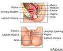
Internal structures of the female reproductive anatomy include the uterus, ovaries, and cervix. External structures include the labium minora and majora, the vagina and the clitoris.
Female reproductive anatomy
illustration
-
Endometrial biopsy - illustration
The mucosal lining of the cavity of the uterus is called the endometrium. It is this lining which undergoes changes over the course of the monthly menstrual cycle, sloughes off and becomes part of the menses. A biopsy of the endometrium is used to check for disease or problems of fertility.
Endometrial biopsy
illustration
-
Hysterectomy - illustration
Hysterectomy is surgical removal of the uterus, resulting in inability to become pregnant. This surgery may be done for a variety of reasons including, but not restricted to, chronic pelvic inflammatory disease, uterine fibroids and cancer. A hysterectomy may be done through an abdominal or a vaginal incision.
Hysterectomy
illustration
-
Uterus - illustration
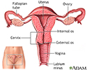
The uterus is a hollow muscular organ located in the female pelvis between the bladder and rectum. The ovaries produce the eggs that travel through the fallopian tubes. Once the egg has left the ovary it can be fertilized and implant itself in the lining of the uterus. The main function of the uterus is to nourish the developing fetus prior to birth.
Uterus
illustration
-
Endometrial cancer - illustration
Endometrial cancer is a cancerous growth of the endometrium (lining of the uterus). It is the most common uterine cancer.
Endometrial cancer
illustration
-
Pelvic laparoscopy - illustration
Laparoscopy is performed when less-invasive surgery is desired. It is also called Band-Aid surgery because only small incisions need to be made to accommodate the small surgical instruments that are used to view the abdominal contents and perform the surgery.
Pelvic laparoscopy
illustration
-
Female reproductive anatomy - illustration
Internal structures of the female reproductive anatomy include the uterus, ovaries, and cervix. External structures include the labium minora and majora, the vagina and the clitoris.
Female reproductive anatomy
illustration
-
Endometrial biopsy - illustration
The mucosal lining of the cavity of the uterus is called the endometrium. It is this lining which undergoes changes over the course of the monthly menstrual cycle, sloughes off and becomes part of the menses. A biopsy of the endometrium is used to check for disease or problems of fertility.
Endometrial biopsy
illustration
-
Hysterectomy - illustration
Hysterectomy is surgical removal of the uterus, resulting in inability to become pregnant. This surgery may be done for a variety of reasons including, but not restricted to, chronic pelvic inflammatory disease, uterine fibroids and cancer. A hysterectomy may be done through an abdominal or a vaginal incision.
Hysterectomy
illustration
-
Uterus - illustration
The uterus is a hollow muscular organ located in the female pelvis between the bladder and rectum. The ovaries produce the eggs that travel through the fallopian tubes. Once the egg has left the ovary it can be fertilized and implant itself in the lining of the uterus. The main function of the uterus is to nourish the developing fetus prior to birth.
Uterus
illustration
-
Endometrial cancer - illustration
Endometrial cancer is a cancerous growth of the endometrium (lining of the uterus). It is the most common uterine cancer.
Endometrial cancer
illustration
Review Date: 3/31/2024
Reviewed By: LaQuita Martinez, MD, Department of Obstetrics and Gynecology, Emory Johns Creek Hospital, Alpharetta, GA. Also reviewed by David C. Dugdale, MD, Medical Director, Brenda Conaway, Editorial Director, and the A.D.A.M. Editorial team.


