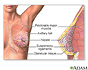
Fibrocystic breasts
Fibrocystic breast disease; Mammary dysplasia; Diffuse cystic mastopathy; Benign breast disease; Glandular breast changes; Cystic changes; Chronic cystic mastitis; Breast lump - fibrocystic; Fibrocystic breast changesFibrocystic breasts are painful, lumpy breasts. Formerly called fibrocystic breast disease, this common condition is, in fact, not a disease. Many women experience these normal breast changes, usually around their period.
Causes
Fibrocystic breast changes occur when thickening of breast tissue (fibrosis) and fluid-filled cysts develop in one or both breasts. It is thought that hormones made in the ovaries during menstruation can trigger these breast changes. This may make your breasts feel swollen, lumpy, or painful before or during your period each month.
More than half of women have this condition at some time during their life. It is most common between the ages of 30 and 50. It is rare in women after menopause unless they are taking estrogen. Fibrocystic breast changes do not change your risk for breast cancer.
Symptoms
Symptoms are more often worse right before your menstrual period. They tend to get better after your period starts.
If you have heavy, irregular periods, your symptoms may be worse. If you take birth control pills, you may have fewer symptoms. In most cases, symptoms get better after menopause.
Symptoms may include:
- Pain or discomfort in both breasts that may come and go with your period, but may also last through the whole month
- Breasts that feel full, swollen, or heavy
- Pain or discomfort under the arms
- Breast lumps that change in size with the menstrual period
You may have a lump in the same area of the breast that becomes larger before each period and returns to its original size afterward. This type of lump moves when it is pushed with your fingers. It does not feel stuck or fixed to the tissue around it. This type of lump is common with fibrocystic breasts.
Exams and Tests
Your health care provider will examine you. This will include a breast exam. Tell your provider if you have noticed any breast changes.
Mammography is performed to screen women to detect early breast cancer when it is more likely to be cured. The recommendations of different expert organizations can differ.
Mammography
A mammogram is an x-ray picture of the breasts. It is used to evaluate some breast symptoms and to find breast cancer in women with no symptoms (cal...

- Mammography is generally recommended for all women starting at age 40, repeated every 1 to 2 years.
- Women with a family history of breast cancer should work with their provider to assess their risk of breast cancer. In some situations, additional testing may be considered.
Mammograms have clear benefit for health by finding work best at finding early stage breast cancer in women ages 40 to 74. It is important to note that all women, including those with dense breasts, should be screened starting at age 40. It is unclear if mammograms benefit women ages 75 and older.
For women under 35, a breast ultrasound may be used to look more closely at breast tissue. You may need further tests if a lump was found during a breast exam or your imaging result was abnormal.
Breast ultrasound
Breast ultrasound is a test that uses sound waves to examine the breasts.

If the lump appears to be a cyst, your provider may aspirate the lump with a needle, which confirms the lump was a cyst and sometimes may improve the symptoms. For other types of lumps, additional imaging may be done. If these exams are normal, but your provider still has concerns about a lump, a biopsy may be performed.
Biopsy
A biopsy is the removal of a small piece of tissue for lab examination.

Treatment
Women who have no symptoms or only mild symptoms do not need treatment.
Your provider may recommend the following self-care measures:
- Take over-the-counter medicine, such as acetaminophen or ibuprofen for pain
- Apply heat or ice on the breast
- Wear a well-fitting supportive bra, such as a sports bra
Some women believe that eating less fat, caffeine, or chocolate helps with their symptoms. There is no clear evidence that these measures help large numbers of women.
Vitamin E, thiamine, magnesium, and evening primrose oil are not harmful in most cases. Studies have not shown these to be helpful. Talk with your provider before taking any medicine or supplement.
For more severe symptoms, your provider may prescribe hormones, such as birth control pills or other medicine. Take the medicine exactly as instructed. Be sure to let your provider know if you have side effects from the medicine.
Surgery is never done to treat this condition. However, a lump that stays the same throughout your menstrual cycle is considered suspicious. In this case, your provider may recommend a core needle biopsy. In this test, a small amount of tissue is removed from the lump and examined under a microscope.
Outlook (Prognosis)
If your breast exams and mammograms are normal, you do not need to worry about your symptoms. Fibrocystic breast changes do not generally increase your risk for breast cancer. However, if you have a family history of breast cancer and fibrocystic changes, there is a small increase in the risk. Symptoms usually improve after menopause.
When to Contact a Medical Professional
Contact your provider if:
- You notice any unfamiliar changes in the appearance or texture of your breasts.
- You have new discharge from the nipple or any discharge that is bloody or clear.
- You have redness or puckering of the skin, or flattening or indentation of the nipple.
References
American College of Obstetricians and Gynecologists website. Benign breast conditions. www.acog.org/womens-health/faqs/benign-breast-problems-and-conditions. Updated January 2023. Accessed November 7, 2024.
Cox DM, Lippe C, Geletzke AK, et al. Etiology and management of benign breast disease. In: Klimberg VS, Gradishar WJ, Bland KI, Korourian S, White J, Copeland EM, eds. Bland and Copeland's The Breast: Comprehensive Management of Benign and Malignant Diseases. 6th ed. Philadelphia, PA: Elsevier; 2024:chap 14.
Klimberg VS, Hunt KK. Diseases of the breast. In: Townsend CM Jr, Beauchamp RD, Evers BM, Mattox KL, eds. Sabiston Textbook of Surgery. 21st ed. St Louis, MO: Elsevier; 2022:chap 35.
Sandadi S, Rock DT, Orr JW, Valea FA. Breast diseases: detection, management, and surveillance of breast disease. In: Gershenson DM, Lentz GM, Valea FA, Lobo RA, eds. Comprehensive Gynecology. 8th ed. Philadelphia, PA: Elsevier; 2022:chap 15.
US Preventive Services Task Force; Nicholson WK, Silverstein M, Wong JB, et al. Screening for breast cancer: US Preventive Services Task Force Recommendation Statement. JAMA. 2024;331(22):1918-1930. PMID: 38687503. pubmed.ncbi.nlm.nih.gov/38687503/.
-
Female Breast - illustration
The female breast is either of two mammary glands (organs of milk secretion) on the chest.
Female Breast
illustration
-
Fibrocystic breast change - illustration
Fibrocystic breast change is a common and benign change within the breast characterized by a dense irregular and bumpy consistency in the breast tissue. Mammography or biopsy may be needed to rule out other disorders.
Fibrocystic breast change
illustration
-
Female Breast - illustration
The female breast is either of two mammary glands (organs of milk secretion) on the chest.
Female Breast
illustration
-
Fibrocystic breast change - illustration
Fibrocystic breast change is a common and benign change within the breast characterized by a dense irregular and bumpy consistency in the breast tissue. Mammography or biopsy may be needed to rule out other disorders.
Fibrocystic breast change
illustration
Review Date: 10/17/2024
Reviewed By: Linda J. Vorvick, MD, Clinical Professor, Department of Family Medicine, UW Medicine, School of Medicine, University of Washington, Seattle, WA. Also reviewed by David C. Dugdale, MD, Medical Director, Brenda Conaway, Editorial Director, and the A.D.A.M. Editorial team.


