Otosclerosis
Otospongiosis; Hearing loss - otosclerosisOtosclerosis is an abnormal bone growth in the middle ear that causes hearing loss.
Hearing loss
Hearing loss is being partly or totally unable to hear sound in one or both ears.

Causes
The exact cause of otosclerosis is unknown. It may be passed down through families.
People who have otosclerosis have an abnormal extension of sponge-like bone growing in the middle ear cavity. This growth prevents the ear bones from vibrating normally in response to sound waves. These vibrations are needed in order for you to hear.
Otosclerosis is the most common cause of middle ear hearing loss in young adults. It typically begins in early to mid-adulthood. It is more common in women than in men. The condition may affect one or both ears.
Risks for this condition include pregnancy and a family history of hearing loss. White people are more likely to develop this condition than people of other races.
Symptoms
Symptoms include:
- Hearing loss (slow at first, but worsens over time)
-
Ringing in the affected ear (tinnitus)
Tinnitus
Tinnitus is the medical term for "hearing" noises in your ears. It occurs when there is no outside source of the sounds. Tinnitus is often called "r...
 ImageRead Article Now Book Mark Article
 - Vertigo or dizziness
Exams and Tests
A hearing test (audiometry/audiology) may help determine the severity of hearing loss.
Audiometry
An audiometry exam tests your ability to hear sounds. Sounds vary, based on their loudness (intensity) and the speed of sound wave vibrations (tone)...

Audiology
An audiometry exam tests your ability to hear sounds. Sounds vary, based on their loudness (intensity) and the speed of sound wave vibrations (tone)...

A special imaging test of the head called a temporal-bone CT may be used to look for other causes of hearing loss.
Treatment
Otosclerosis may slowly get worse. The condition may not need to be treated until you have more serious hearing problems.
Using some medicines such as fluoride, calcium, or vitamin D may help to slow the hearing loss. However, the benefits of these treatments have not yet been proven.
Vitamin D
Vitamin D is a fat-soluble vitamin. Fat-soluble vitamins are stored in the body's fatty tissue and liver.

A hearing aid may be used to treat the hearing loss. This will not cure or prevent hearing loss from getting worse, but it may help with symptoms.
Surgery can cure or improve conductive hearing loss. Either all or part of one of the small middle ear bones behind the eardrum (stapes) is removed and replaced with a prosthesis.
Prosthesis
A prosthesis is a device designed to replace a missing part of the body or to make a part of the body work better. Diseased or missing eyes, arms, h...

- A total replacement is called a stapedectomy.
- Sometimes only part of the stapes is removed and a small hole is made in the bottom of it. This is called a stapedotomy. Sometimes a laser is used to help with the surgery.
Outlook (Prognosis)
Otosclerosis gets worse without treatment. Surgery can restore some or all of your hearing loss. Pain and dizziness from the surgery go away within a few weeks for most people.
To reduce the risk of complications after surgery:
- DO NOT blow your nose for 2 to 3 weeks after surgery.
- Avoid people with respiratory or other infections.
Respiratory
The words "respiratory" and "respiration" refer to the lungs and breathing.
 ImageRead Article Now Book Mark Article
 - Avoid bending, lifting, or straining, which may cause dizziness.
Dizziness.
Dizziness is a term that is often used to describe 2 different symptoms: lightheadedness and vertigo. Lightheadedness is a feeling that you might fai...
 ImageRead Article Now Book Mark Article
 - Avoid loud noises or sudden pressure changes, such as scuba diving, flying, or driving in the mountains until you have healed.
If surgery does not work, you may have total hearing loss. Treatment for total hearing loss involves developing skills to cope with deafness, and using hearing aids to transmit sounds from the non-hearing ear to the good ear.
Deafness
Hearing loss is being partly or totally unable to hear sound in one or both ears.

Possible Complications
Complications may include:
- Complete deafness
- Abnormal taste in the mouth or loss of taste to part of the tongue, temporary or permanent
- Infection, dizziness, pain, or a blood clot in the ear after surgery
Blood clot
Blood clots are clumps that occur when blood hardens from a liquid to a solid. A blood clot that forms inside one of your veins or arteries is calle...
 ImageRead Article Now Book Mark Article
 - Nerve damage
When to Contact a Medical Professional
Contact your health care provider if:
- You have hearing loss
- You develop fever, ear pain, dizziness, or other symptoms after surgery
References
House JW, Cunningham CD. Otosclerosis. In: Flint PW, Francis HW, Haughey BH, et al, eds. Cummings Otolaryngology: Head and Neck Surgery. 7th ed. Philadelphia, PA: Elsevier; 2021:chap 146.
Ironside JW, Smith C. Central and peripheral nervous systems. In: Cross SS, ed. Underwood Pathology. 7th ed. Philadelphia, PA: Elsevier; 2019:chap 26.
O'Handley JG, Tobin EJ, Shah AR. Otorhinolaryngology. In: Rakel RE, Rakel DP, eds. Textbook of Family Medicine. 9th ed. Philadelphia, PA: Elsevier; 2016:chap 18.
Rivero A, Yoshikawa N. Otosclerosis. In: Myers EN, Snyderman CH, eds. Operative Otolaryngology Head and Neck Surgery. 3rd ed. Philadelphia, PA: Elsevier; 2018:chap 133.
-
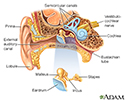
Ear anatomy - illustration
The ear consists of external, middle, and inner structures. The eardrum and the 3 tiny bones conduct sound from the eardrum to the cochlea.
Ear anatomy
illustration
Review Date: 5/2/2024
Reviewed By: Josef Shargorodsky, MD, MPH, Johns Hopkins University School of Medicine, Baltimore, MD. Also reviewed by David C. Dugdale, MD, Medical Director, Brenda Conaway, Editorial Director, and the A.D.A.M. Editorial team.


