Endocarditis
Valve infection; Staphylococcus aureus - endocarditis; Enterococcus - endocarditis; Streptococcus viridans - endocarditis; Candida - endocarditisEndocarditis is inflammation of the inside lining of the heart chambers and heart valves (endocardium). It is most often caused by a bacterial or, rarely, a fungal infection.
Causes
Endocarditis can involve the heart muscle, heart valves, or lining of the heart. Some people who develop endocarditis have a:
-
Birth defect of the heart
Birth defect of the heart
Congenital heart disease (CHD) is a problem with the heart's structure and function that is present at birth.
 ImageRead Article Now Book Mark Article
 - Damaged or abnormal heart valve
- History of endocarditis
- New heart valve after surgery
- Parenteral (intravenous) drug use disorder or addiction
- Long-term intravenous line in place
Endocarditis begins when germs enter the bloodstream and then travel to the heart.
- Bacterial infection is the most common cause of endocarditis.
- Endocarditis can also be caused by fungi, such as Candida.
- In some cases, no cause can be found.
Germs are most likely to enter the bloodstream during:
- Central venous access lines
- Injection drug use, from the use of unclean (unsterile) needles
- Recent dental surgery
- Other surgeries or minor procedures to the breathing tract, urinary tract, infected skin, or bones and muscles
Symptoms
Symptoms of endocarditis may develop slowly or suddenly.
Fever, chills, and sweating are frequent symptoms. These sometimes can:
Fever
Fever is the temporary increase in the body's temperature in response to a disease or illness. A child has a fever when the temperature is at or abov...

Chills
Chills refers to feeling cold after being in a cold environment. The word can also refer to an episode of shivering along with paleness and feeling ...

Sweating
Sweating is the release of liquid from the body's sweat glands. This liquid contains salt. This process is also called perspiration. Sweating helps...

- Be present for days before any other symptoms appear
- Come and go, or be more noticeable at nighttime
You may also have fatigue, weakness, weight loss, loss of appetite, and aches and pains in the muscles or joints.
Fatigue
Fatigue is a feeling of weariness, tiredness, or lack of energy.

Muscles
Muscle aches and pains are common and can involve more than one muscle. Pain due to muscles can also be felt in ligaments, tendons, and fascia. Fas...

Other signs can include:
- Small areas of bleeding under the nails (splinter hemorrhages)
Splinter hemorrhages
Splinter hemorrhages are small areas of bleeding (hemorrhage) under the fingernails or toenails.
 ImageRead Article Now Book Mark Article
 - Red, painless skin spots on the palms and soles (Janeway lesions)
- Red, painful nodes in the pads of the fingers and toes (Osler nodes)
-
Shortness of breath with activity
Shortness of breath
Breathing difficulty may involve:Difficult breathing Uncomfortable breathingFeeling like you are not getting enough air
 ImageRead Article Now Book Mark Article
 - Swelling of feet, legs, abdomen
Exams and Tests
The health care provider may detect a new heart murmur, or a change in an existing heart murmur.
Heart murmur
A heart murmur is a blowing, whooshing, or rasping sound heard during a heartbeat. The sound is caused by turbulent (rough) blood flow through the h...

An eye exam may show bleeding in the retina and a central area of clearing. This finding is known as Roth spots. There may be small, pinpoint areas of bleeding on the surface of the eye or the eyelids called petechiae.
Tests that may be done include:
-
Blood culture to identify the bacteria or fungus that is causing the infection
Blood culture
A blood culture is a laboratory test to check for bacteria or other germs in a blood sample.
 ImageRead Article Now Book Mark Article
 -
Complete blood count (CBC), C-reactive protein (CRP), or erythrocyte sedimentation rate (ESR)
Complete blood count
A complete blood count (CBC) test measures the following:The number of white blood cells (WBC count)The number of red blood cells (RBC count)The numb...
 ImageRead Article Now Book Mark Article
Erythrocyte sedimentation rate
ESR stands for erythrocyte sedimentation rate. It is commonly called a "sed rate. "It is a test that indirectly measures the level of certain protei...
 ImageRead Article Now Book Mark Article
 - An echocardiogram to look at the heart valves
Echocardiogram
An echocardiogram is a test that uses sound waves to create pictures of the heart. The picture and information it produces is more detailed than a s...
 ImageRead Article Now Book Mark Article
Treatment
You may need to be in the hospital to get antibiotics through a vein (IV or intravenously). Blood cultures and tests will help your provider choose the best antibiotic.
You will then need long-term antibiotic therapy.
- People most often need therapy for 4 to 6 weeks to kill all the bacteria from the heart chambers and valves.
- Antibiotic treatments that are started in the hospital will need to be continued at home.
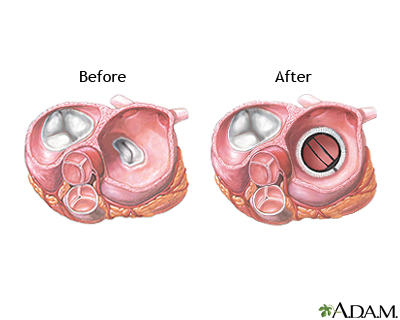
Surgery to replace the heart valve is often needed when:
- The infection is breaking off into little pieces, resulting in strokes or blockages of other arteries.
- The person develops heart failure as a result of damaged heart valves.
- There is evidence of more severe organ damage (such as heart damage).
Outlook (Prognosis)
Getting treatment for endocarditis right away improves the chances of a good outcome.
More serious problems that may develop include:
-
Brain abscess
Brain abscess
A brain abscess is a collection of pus, immune cells, and other material in the brain, caused by a bacterial or fungal infection.
 ImageRead Article Now Book Mark Article
 - Further damage to the heart valves, causing heart failure
Heart failure
Heart failure is a condition in which the heart is no longer able to pump oxygen-rich blood to the rest of the body efficiently. This causes symptom...
 ImageRead Article Now Book Mark Article
 - Spread of the infection to other parts of the body
-
Stroke, caused by small clots or pieces of the infection breaking off and traveling to the brain
Stroke
A stroke occurs when blood flow to a part of the brain stops. A stroke is sometimes called a "brain attack. " If blood flow is cut off for longer th...
 ImageRead Article Now Book Mark Article
When to Contact a Medical Professional
Contact your provider if you notice the following symptoms during or after treatment:
- Blood in urine
-
Chest pain
Chest pain
Chest pain is discomfort or pain that you feel anywhere along the front of your body between your neck and upper abdomen.
 ImageRead Article Now Book Mark Article
 - Fatigue
- Fever that doesn't go away
- Fever
-
Numbness
Numbness
Numbness and tingling are abnormal sensations that can occur anywhere in your body, but they are often felt in your fingers, hands, feet, arms, or le...
 ImageRead Article Now Book Mark Article
 - Weakness
-
Weight loss without change in diet
Weight loss
Unexplained weight loss is a decrease in body weight, when you did not try to lose the weight on your own. Many people gain and lose weight. Uninten...
Read Article Now Book Mark Article
Prevention
The American Heart Association recommends preventive antibiotics for people at risk for infectious endocarditis, such as those with:
- Certain birth defects of the heart
Birth defects of the heart
Congenital heart defect corrective surgery fixes or treats a heart defect that a child is born with. A baby born with one or more heart defects has ...
 ImageRead Article Now Book Mark Article
 -
Heart transplant and valve problems
Heart transplant
A heart transplant is surgery to remove a damaged or diseased heart and replace it with a healthy donor heart.
 ImageRead Article Now Book Mark Article
 - Prosthetic heart valves (heart valves inserted by a surgeon)
- Past history of endocarditis
These people should receive antibiotics when they have:
- Dental procedures that are likely to cause bleeding
- Procedures involving the breathing tract
- Procedures involving the urinary tract
- Procedures involving the digestive tract
- Procedures on skin infections and soft tissue infections
References
Baddour LM, Anavekar NS, Crestanello JA, Wilson WR. Infectious endocarditis and infections of indwelling devices. In: Libby P, Bonow RO, Mann DL, Tomaselli GF, Bhatt DL, Solomon SD, eds. Braunwald's Heart Disease: A Textbook of Cardiovascular Medicine. 12th ed. Philadelphia, PA: Elsevier; 2022:chap 80.
Fowler VG, Bayer AS, Baddour LM. Infective endocarditis. In: Goldman L, Cooney KA, eds. Goldman-Cecil Medicine. 27th ed. Philadelphia, PA: Elsevier; 2024:chap 61.
Holland TL, Bayer AS, Fowler VG. Endocarditis and intravascular infections. In: Bennett JE, Dolin R, Blaser MJ, eds. Mandell, Douglas, and Bennett's Principles and Practice of Infectious Diseases. 9th ed. Philadelphia, PA: Elsevier; 2020:chap 80.
-
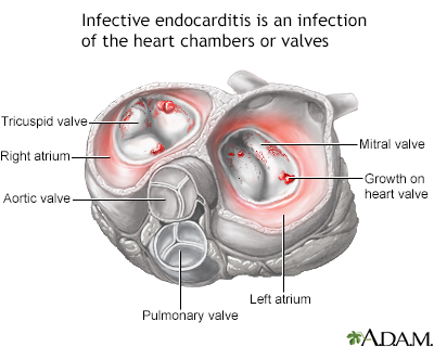
Infective endocarditis - illustration
Infectious endocarditis involves the heart valves and is most commonly found in people who have underlying heart disease. Sources of the infection may be transient bacteremia, which is common during dental, upper respiratory, urologic, and lower gastrointestinal diagnostic and surgical procedures. The infection can cause growths on the heart valves, the lining of the heart, or the lining of the blood vessels. These growths may be dislodged and send clots to the brain, lungs, kidneys, or spleen.
Infective endocarditis
illustration
-
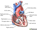
Heart - section through the middle - illustration
The interior of the heart is composed of valves, chambers, and associated vessels.
Heart - section through the middle
illustration
-
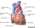
Heart - front view - illustration
The external structures of the heart include the ventricles, atria, arteries and veins. Arteries carry blood away from the heart while veins carry blood into the heart. The vessels colored blue indicate the transport of blood with relatively low content of oxygen and high content of carbon dioxide. The vessels colored red indicate the transport of blood with relatively high content of oxygen and low content of carbon dioxide.
Heart - front view
illustration
-
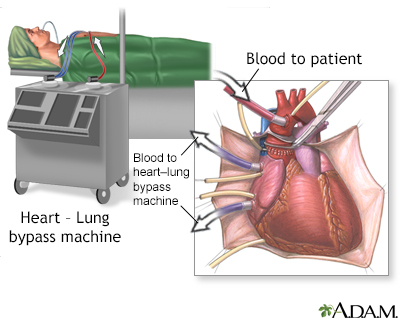
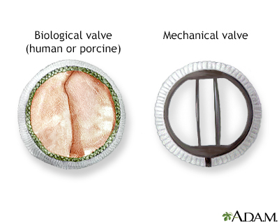
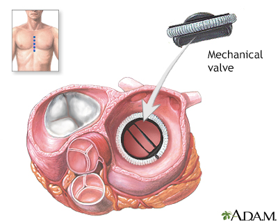
Heart valve surgery - Series
Presentation
-
Janeway lesion - close-up - illustration
Janeway lesions are seen in people with acute bacterial endocarditis. They appear as flat, painless, red to bluish-red spots on the palms and soles.
Janeway lesion - close-up
illustration
-
Janeway lesion on the finger - illustration
Janeway lesions appear as flat, painless, red or reddish-blue patches on the hands and soles of people with acute bacterial endocarditis.
Janeway lesion on the finger
illustration
-
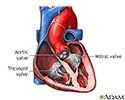
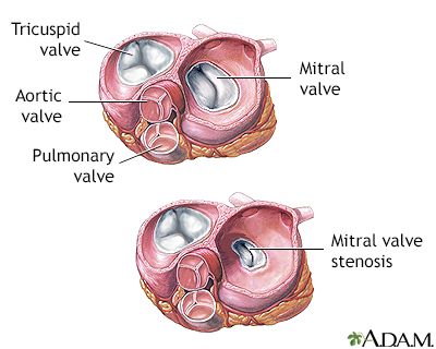
Heart valves - illustration
The valves of the heart open and close to control the flow of blood entering or leaving the heart.
Heart valves
illustration
-
Infective endocarditis - illustration
Infectious endocarditis involves the heart valves and is most commonly found in people who have underlying heart disease. Sources of the infection may be transient bacteremia, which is common during dental, upper respiratory, urologic, and lower gastrointestinal diagnostic and surgical procedures. The infection can cause growths on the heart valves, the lining of the heart, or the lining of the blood vessels. These growths may be dislodged and send clots to the brain, lungs, kidneys, or spleen.
Infective endocarditis
illustration
-
Heart - section through the middle - illustration
The interior of the heart is composed of valves, chambers, and associated vessels.
Heart - section through the middle
illustration
-
Heart - front view - illustration
The external structures of the heart include the ventricles, atria, arteries and veins. Arteries carry blood away from the heart while veins carry blood into the heart. The vessels colored blue indicate the transport of blood with relatively low content of oxygen and high content of carbon dioxide. The vessels colored red indicate the transport of blood with relatively high content of oxygen and low content of carbon dioxide.
Heart - front view
illustration
-
Heart valve surgery - Series
Presentation
-
Janeway lesion - close-up - illustration
Janeway lesions are seen in people with acute bacterial endocarditis. They appear as flat, painless, red to bluish-red spots on the palms and soles.
Janeway lesion - close-up
illustration
-
Janeway lesion on the finger - illustration
Janeway lesions appear as flat, painless, red or reddish-blue patches on the hands and soles of people with acute bacterial endocarditis.
Janeway lesion on the finger
illustration
-
Heart valves - illustration
The valves of the heart open and close to control the flow of blood entering or leaving the heart.
Heart valves
illustration
Review Date: 11/10/2024
Reviewed By: Jatin M. Vyas, MD, PhD, Roy and Diana Vagelos Professor in Medicine, Columbia University Vagelos College of Physicians and Surgeons, Division of Infectious Diseases, Department of Medicine, New York, NY. Also reviewed by David C. Dugdale, MD, Medical Director, Brenda Conaway, Editorial Director, and the A.D.A.M. Editorial team.

