Congenital heart disease
Congenital heart disease (CHD) is a problem with the heart's structure and function that is present at birth.
Causes
CHD can describe a number of different problems affecting the heart. It is the most common type of birth defect. CHD causes more deaths in the first year of life than any other birth defects.
CHD is often divided into two types: cyanotic (blue skin color caused by a lack of oxygen) and non-cyanotic. The following lists cover the most common CHDs:
Cyanotic
A bluish color to the skin or mucous membrane is usually due to a lack of oxygen in the blood. The medical term is cyanosis.

Cyanotic:
-
Ebstein anomaly
Ebstein anomaly
Ebstein anomaly is a rare heart defect in which parts of the tricuspid valve are abnormal. The tricuspid valve separates the right lower heart chamb...
 ImageRead Article Now Book Mark Article
 -
Hypoplastic left heart
Hypoplastic left heart
Hypoplastic left heart syndrome occurs when parts of the left side of the heart (mitral valve, left ventricle, aortic valve, and aorta) do not develo...
 ImageRead Article Now Book Mark Article
 -
Pulmonary atresia
Pulmonary atresia
Pulmonary atresia is a form of heart disease in which the pulmonary valve does not form properly. It is present from birth (congenital heart disease...
 ImageRead Article Now Book Mark Article
 -
Tetralogy of Fallot
Tetralogy of Fallot
Tetralogy of Fallot is a type of congenital heart defect. Congenital means that it is present at birth.
 ImageRead Article Now Book Mark Article
 -
Total anomalous pulmonary venous return
Total anomalous pulmonary venous return
Total anomalous pulmonary venous return (TAPVR) is a heart disease in which the 4 veins that take blood from the lungs to the heart do not attach nor...
 ImageRead Article Now Book Mark Article
 -
Transposition of the great vessels
Transposition of the great vessels
Transposition of the great arteries (TGA) is a heart defect that occurs from birth (congenital). The two major arteries that carry blood away from t...
 ImageRead Article Now Book Mark Article
 -
Tricuspid atresia
Tricuspid atresia
Tricuspid atresia is a type of heart disease that is present at birth (congenital heart disease), in which the tricuspid heart valve is missing or ab...
 ImageRead Article Now Book Mark Article
 -
Truncus arteriosus
Truncus arteriosus
Truncus arteriosus is a rare type of heart disease in which a single blood vessel (truncus arteriosus) comes out of the right and left ventricles, in...
 ImageRead Article Now Book Mark Article
Non-cyanotic:
-
Aortic stenosis
Aortic stenosis
The aorta is the main artery that carries blood out of the heart to the rest of the body. Blood flows out of the heart and into the aorta through th...
 ImageRead Article Now Book Mark Article
 - Bicuspid aortic valve
-
Atrial septal defect (ASD)
Atrial septal defect
Atrial septal defect (ASD) is a heart defect that is present at birth (congenital). As a baby develops in the womb, a wall (septum) forms that divide...
 ImageRead Article Now Book Mark Article
 - Atrioventricular canal (endocardial cushion defect)
Endocardial cushion defect
Endocardial cushion defect (ECD) is an abnormal heart condition. The walls separating all four chambers of the heart are poorly formed or absent. A...
 ImageRead Article Now Book Mark Article
 -
Coarctation of the aorta
Coarctation of the aorta
The aorta is a large artery that carries blood from the heart to the vessels that supply the rest of the body with blood. If part of the aorta is na...
 ImageRead Article Now Book Mark Article
 -
Patent ductus arteriosus (PDA)
Patent ductus arteriosus
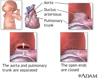
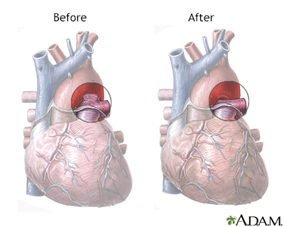
Patent ductus arteriosus (PDA) is a condition in which the ductus arteriosus does not close. The word "patent" means open. The ductus arteriosus is ...
 ImageRead Article Now Book Mark Article
 -
Pulmonic stenosis
Pulmonic stenosis
Pulmonic stenosis is a heart valve disorder that involves the pulmonary valve. This is the valve separating the right ventricle (one of the chambers ...
 ImageRead Article Now Book Mark Article
 -
Ventricular septal defect (VSD)
Ventricular septal defect
A ventricular septal defect is a hole in the wall that separates the right and left ventricles of the heart. Ventricular septal defect is one of the...
 ImageRead Article Now Book Mark Article
These problems may occur alone or together. Most children with CHD do not have other types of birth defects. However, heart defects may be part of genetic and chromosomal syndromes. Some of these syndromes may be passed down through families.
Examples include:
- DiGeorge syndrome
-
Down syndrome
Down syndrome
Down syndrome is a genetic condition in which a person has 47 chromosomes instead of the usual 46.
Read Article Now Book Mark Article -
Marfan syndrome
Marfan syndrome
Marfan syndrome is a disorder of connective tissue. This is the tissue that strengthens the body's structures. Disorders of connective tissue affect...
 ImageRead Article Now Book Mark Article
 -
Noonan syndrome
Noonan syndrome
Noonan syndrome is a disease present from birth (congenital) that causes many parts of the body to develop abnormally. In about 50% of cases, it is ...
 ImageRead Article Now Book Mark Article
 -
Edwards syndrome
Edwards syndrome
Trisomy 18 is a genetic disorder in which a person has a third copy of material from chromosome 18, instead of the usual 2 copies. Rarely, the extra...
Read Article Now Book Mark Article -
Trisomy 13
Trisomy 13
Trisomy 13 (also called Patau syndrome) is a genetic disorder in which a person has 3 copies of genetic material from chromosome 13, instead of the u...
 ImageRead Article Now Book Mark Article
 -
Turner syndrome
Turner syndrome
Turner syndrome is a rare genetic condition in which a female does not have the usual pair of X chromosomes.
 ImageRead Article Now Book Mark Article
Often, no cause for the heart disease can be found. CHDs continue to be investigated and researched. Medicines such as retinoic acid for acne, chemicals, alcohol, and infections (such as rubella) during pregnancy can contribute to some congenital heart problems.
Rubella
Rubella, also known as the German measles, is an infection in which there is a rash on the skin. Congenital rubella is when a pregnant woman with rub...

Poorly controlled blood sugar in women who have diabetes during pregnancy has also been linked to an increased rate of congenital heart defects.
Symptoms
Symptoms depend on the condition. Although CHD is present at birth, the symptoms may not appear right away.
Defects such as coarctation of the aorta may not cause problems for years. Other problems, such as a small VSD, ASD, or PDA may never cause any problems.
Coarctation of the aorta
The aorta is a large artery that carries blood from the heart to the vessels that supply the rest of the body with blood. If part of the aorta is na...

Exams and Tests
Most congenital heart defects are found during a pregnancy ultrasound. When a defect is found this way, a pediatric heart doctor, surgeon, and other specialists can be there when the baby is delivered. Having medical care ready at the delivery can mean the difference between life and death for some babies.
Pregnancy ultrasound
A pregnancy ultrasound is an imaging test that uses sound waves to create a picture of how a baby is developing in the womb (uterus). It is also use...

Which tests are done on the baby depend on the defect and the symptoms.
Treatment
Which treatment is used, and how well the baby responds to it, depends on the condition. Many defects need to be followed carefully. Some will heal over time, while others will need to be treated.
Some CHDs can be treated with medicine alone. Others need to be treated with one or more heart procedures or surgeries.
Prevention
Women who plan to become pregnant should be immunized against rubella if they are not already immune. Rubella infection in a pregnant woman can cause CHD.
Women who are pregnant should get good prenatal care:
- Avoid alcohol and illegal drugs during pregnancy.
- Tell your health care provider that you are pregnant before taking any new medicines.
- Have a blood test early in your pregnancy to see if you are immune to rubella. If you are not immune, avoid any possible exposure to rubella and get vaccinated right after delivery.
- Pregnant women who have diabetes should try to get good control over their blood sugar level.
Good control over their blood sugar lev...
Gestational diabetes is diabetes, defined by high blood sugar (glucose), that starts during pregnancy. If you've been diagnosed with gestational dia...
 ImageRead Article Now Book Mark Article
Certain genes may play a role in CHD. Many family members may be affected. Talk to your provider about genetic counseling and screening if you have a family history of CHD.
References
Bernstein D. Evaluation and screening of the infant or child with congenital heart disease. In: Kliegman RM, St. Geme JW, Blum NJ, et al, eds. Nelson Textbook of Pediatrics. 22nd ed. Philadelphia, PA: Elsevier; 2025:chap 474.
Valente AM, Dorfman AL, Babu-Narayan SV, Kreiger EV. Congenital heart disease in the adolescent and adult. In: Libby P, Bonow RO, Mann DL, Tomaselli GF, Bhatt DL, Solomon SD, eds. Braunwald's Heart Disease: A Textbook of Cardiovascular Medicine. 12th ed. Philadelphia, PA: Elsevier; 2022:chap 82.
Well A, Fraser CD. Congenital heart disease. In: Townsend CM Jr, Beauchamp RD, Evers BM, Mattox KL, eds. Sabiston Textbook of Surgery. 21st ed. St Louis, MO: Elsevier; 2022:chap 59.
-
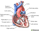
Heart - section through the middle - illustration
The interior of the heart is composed of valves, chambers, and associated vessels.
Heart - section through the middle
illustration
-
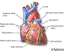
Heart - front view - illustration
The external structures of the heart include the ventricles, atria, arteries and veins. Arteries carry blood away from the heart while veins carry blood into the heart. The vessels colored blue indicate the transport of blood with relatively low content of oxygen and high content of carbon dioxide. The vessels colored red indicate the transport of blood with relatively high content of oxygen and low content of carbon dioxide.
Heart - front view
illustration
-
Ultrasound, normal fetus - heartbeat - illustration
This is a normal fetal ultrasound showing one pattern of the fetal heartbeat. Some ultrasound machines have the ability to focus on different areas of the heart and evaluate the heartbeat. This is useful in the early diagnosis of congenital heart abnormalities.
Ultrasound, normal fetus - heartbeat
illustration
-
Ultrasound, ventricular septal defect - heartbeat - illustration
This is an ultrasound showing a ventricular septal defect pattern of the fetal heartbeat. Some ultrasound machines have the ability to focus on different areas of the heart and evaluate the heartbeat. This is useful in the early diagnosis of congenital heart abnormalities.
Ultrasound, ventricular septal defect - heartbeat
illustration
-
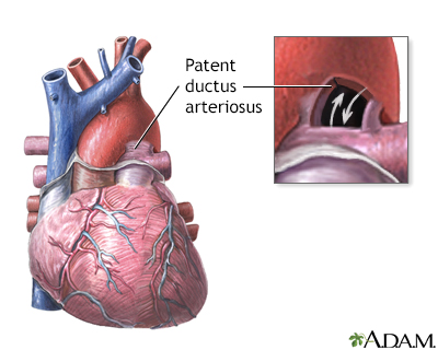
Patent ductus arteriosis (PDA) - series - Infant heart anatomy
Presentation
-
Heart - section through the middle - illustration
The interior of the heart is composed of valves, chambers, and associated vessels.
Heart - section through the middle
illustration
-
Heart - front view - illustration
The external structures of the heart include the ventricles, atria, arteries and veins. Arteries carry blood away from the heart while veins carry blood into the heart. The vessels colored blue indicate the transport of blood with relatively low content of oxygen and high content of carbon dioxide. The vessels colored red indicate the transport of blood with relatively high content of oxygen and low content of carbon dioxide.
Heart - front view
illustration
-
Ultrasound, normal fetus - heartbeat - illustration
This is a normal fetal ultrasound showing one pattern of the fetal heartbeat. Some ultrasound machines have the ability to focus on different areas of the heart and evaluate the heartbeat. This is useful in the early diagnosis of congenital heart abnormalities.
Ultrasound, normal fetus - heartbeat
illustration
-
Ultrasound, ventricular septal defect - heartbeat - illustration
This is an ultrasound showing a ventricular septal defect pattern of the fetal heartbeat. Some ultrasound machines have the ability to focus on different areas of the heart and evaluate the heartbeat. This is useful in the early diagnosis of congenital heart abnormalities.
Ultrasound, ventricular septal defect - heartbeat
illustration
-
Patent ductus arteriosis (PDA) - series - Infant heart anatomy
Presentation
Review Date: 10/1/2025
Reviewed By: Thomas S. Metkus MD, PhD, Associate Professor of Medicine and Surgery, Johns Hopkins University School of Medicine, Baltimore, MD. Also reviewed by David C. Dugdale, MD, Medical Director, Brenda Conaway, Editorial Director, and the A.D.A.M. Editorial team.

