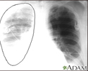
Thoracic aortic aneurysm
Aortic aneurysm - thoracic; Syphilitic aneurysm; Aneurysm - thoracic aorticAn aneurysm is an abnormal widening or ballooning of a portion of an artery due to weakness in the wall of the blood vessel.
A thoracic aortic aneurysm occurs in the part of the body's largest artery (the aorta) that passes through the chest.
Causes
The most common cause of a thoracic aortic aneurysm is hardening of the arteries (atherosclerosis). This condition is more common in people with high cholesterol, long-term high blood pressure, or who smoke.
Hardening of the arteries
Atherosclerosis, sometimes called "hardening of the arteries," occurs when fat, cholesterol, and other substances build up in the walls of arteries. ...

Other risk factors for a thoracic aneurysm include:
- Changes caused by age
- Connective tissue disorders such as Marfan or Ehlers-Danlos syndrome
Marfan
Marfan syndrome is a disorder of connective tissue. This is the tissue that strengthens the body's structures. Disorders of connective tissue affect...
 ImageRead Article Now Book Mark Article
 - Inflammation of the aorta
- Injury from falls or motor vehicle accidents
- Syphilis
Symptoms
Aneurysms develop slowly over many years. Most people have no symptoms until the aneurysm begins to leak or expand.
Symptoms often begin suddenly when:
- The aneurysm grows quickly.
- The aneurysm tears open (called a rupture).
- Blood leaks along the wall of the aorta (aortic dissection).
If the aneurysm presses on nearby structures, the following symptoms may occur:
- Hoarseness
- Swallowing problems
- High-pitched breathing (stridor)
- Swelling in the neck
Other symptoms may include:
- Chest or upper back pain
- Clammy skin
- Nausea and vomiting
- Rapid heart rate
- Sense of impending doom
Exams and Tests
The physical exam is often normal unless a rupture or leak has occurred.
Most thoracic aortic aneurysms are detected on imaging tests performed for other reasons. These tests include chest x-ray, echocardiogram, or chest CT scan or MRI. A chest CT scan shows the size of the aorta and the exact location of the aneurysm.
Chest CT scan
A chest CT (computed tomography) scan is an imaging method that uses x-rays to create cross-sectional pictures of the chest and upper abdomen....

An aortogram (a special set of x-ray images made when dye is injected into the aorta) can identify the aneurysm and any branches of the aorta that may be involved.
Treatment
There is a risk that the aneurysm may open up (rupture) if you do not have surgery to repair it.
The treatment depends on the location of the aneurysm. The aorta is made of three parts:
- The first part moves upward toward the head. It is called the ascending aorta.
- The middle part is curved. It is called the aortic arch.
- The last part moves downward, toward the feet. It is called the descending aorta.
For people with aneurysms of the ascending aorta or aortic arch:
- Surgery to replace the aorta is recommended if an aneurysm is larger than 5 to 6 centimeters (approximately 2 inches).
- A cut is made in the middle of the breast bone (sternum).
- The aorta is replaced with a plastic or fabric graft.
- This is major surgery that requires a heart-lung machine.
For people with aneurysms of the descending thoracic aorta:
- Major surgery is done to replace the aorta with a fabric graft if the aneurysm is larger than 6 centimeters (2.3 inches).
- This surgery is done through a cut on the left side of the chest, which may reach to the abdomen.
- Endovascular stenting is a less invasive option. A stent is a tiny metal or plastic tube that is used to hold an artery open. Stents can be placed into the body without cutting the chest. However, not all people with descending thoracic aneurysms are candidates for stenting.
Stent
A stent is a tiny tube placed into a hollow structure in your body. This structure can be an artery, a vein, or another structure, such as the tube ...
 ImageRead Article Now Book Mark Article
Outlook (Prognosis)
The long-term outlook for people with thoracic aortic aneurysm depends on other medical problems, such as heart disease, high blood pressure, and diabetes. These problems may have caused or contributed to the condition.
Possible Complications
Serious complications after aortic surgery can include:
- Bleeding
- Graft infection
- Heart attack
- Irregular heartbeat
- Kidney damage
- Paralysis
- Stroke
Death soon after the operation occurs in 5% to 10% of people.
Complications after aneurysm stenting include damage to the blood vessels supplying the leg, which may require another operation.
When to Contact a Medical Professional
Contact your health care provider if you have:
- A family history of connective tissue disorders (such as Marfan or Ehlers-Danlos syndrome)
- Chest or back discomfort
Prevention
To prevent atherosclerosis:
- Control your blood pressure and blood lipid levels.
- DO NOT smoke.
- Eat a healthy diet.
- Exercise regularly.
References
Acher CW, Wynn M. Thoracic and thoracoabdominal aneurysms: open surgical treatment. In: Sidawy AN, Perler BA, eds. Rutherford's Vascular Surgery and Endovascular Therapy. 10th ed. Philadelphia, PA: Elsevier; 2023:chap 79.
Beckman JA. Diseases of the aorta. In: Goldman L, Cooney KA, eds. Goldman-Cecil Medicine. 27th ed. Philadelphia, PA: Elsevier; 2024:chap 63.
Braverman AC, Schermerhorn M. Diseases of the aorta. In: Libby P, Bonow RO, Mann DL, Tomaselli GF, Bhatt DL, Solomon SD, eds. Braunwald's Heart Disease: A Textbook of Cardiovascular Medicine. 12th ed. Philadelphia, PA: Elsevier; 2022:chap 42.
Singh MJ, Makaroun MS. Thoracic and thoracoabdominal aneurysms: endovascular treatment. In: Sidawy AN, Perler BA, eds. Rutherford's Vascular Surgery and Endovascular Therapy. 10th ed. Philadelphia, PA: Elsevier; 2023:chap 80.
-
Aortic aneurysm - illustration
Abdominal aortic aneurysm involves a widening, stretching, or ballooning of the aorta. There are several causes of abdominal aortic aneurysm, but the most common results from atherosclerotic disease. As the aorta gets progressively larger over time there is increased chance of rupture.
Aortic aneurysm
illustration
-
Aortic rupture - chest X-ray - illustration
Aortic rupture (a tear in the aorta, which is the major artery coming from the heart) can be seen on a chest X-ray. In this case, it was caused by a traumatic perforation of the thoracic aorta. This is how the X-ray appears when the chest is full of blood (right-sided hemothorax) seen here as cloudiness on the left side of the picture.
Aortic rupture - chest X-ray
illustration
-
Aortic aneurysm - illustration
Abdominal aortic aneurysm involves a widening, stretching, or ballooning of the aorta. There are several causes of abdominal aortic aneurysm, but the most common results from atherosclerotic disease. As the aorta gets progressively larger over time there is increased chance of rupture.
Aortic aneurysm
illustration
-
Aortic rupture - chest X-ray - illustration
Aortic rupture (a tear in the aorta, which is the major artery coming from the heart) can be seen on a chest X-ray. In this case, it was caused by a traumatic perforation of the thoracic aorta. This is how the X-ray appears when the chest is full of blood (right-sided hemothorax) seen here as cloudiness on the left side of the picture.
Aortic rupture - chest X-ray
illustration
Review Date: 5/10/2024
Reviewed By: Neil Grossman, MD, Saint Vincent Radiological Associates, Framingham, MA. Review provided by VeriMed Healthcare Network. Also reviewed by David C. Dugdale, MD, Medical Director, Brenda Conaway, Editorial Director, and the A.D.A.M. Editorial team.


