Congenital cataract
Cataract - congenitalA congenital cataract is a clouding of the lens of the eye that is present at birth. The lens of the eye is normally clear. It focuses light that comes into the eye onto the retina.
Retina
The retina is the light-sensitive layer of tissue at the back of the eyeball. Images that come through the eye's lens are focused on the retina. Th...

Causes
Unlike most cataracts, which occur with aging, congenital cataracts are present at birth.
Congenital cataracts are rare. In most people, no cause can be found.
Congenital cataracts often occur as part of the following birth defects:
- Chondrodysplasia syndrome
- Congenital rubella
- Conradi-Hünermann syndrome
- Down syndrome (trisomy 21)
-
Ectodermal dysplasia syndrome
Ectodermal dysplasia
Ectodermal dysplasias is a group of conditions in which there is abnormal development of the skin, hair, nails, teeth, or sweat glands.
 ImageRead Article Now Book Mark Article
 - Familial congenital cataracts
-
Galactosemia
Galactosemia
Galactosemia is a condition in which the body is unable to use (metabolize) the simple sugar galactose.
 ImageRead Article Now Book Mark Article
 - Hallermann-Streiff syndrome
- Lowe syndrome
- Marinesco-Sjögren syndrome
- Pierre-Robin syndrome
-
Trisomy 13
Trisomy 13
Trisomy 13 (also called Patau syndrome) is a genetic disorder in which a person has 3 copies of genetic material from chromosome 13, instead of the u...
 ImageRead Article Now Book Mark Article
Symptoms
Congenital cataracts most often look different than other forms of cataract.
Symptoms may include:
- An infant does not seem to be visually aware of the world around them (if cataracts are in both eyes)
- Gray or white cloudiness of the pupil (which is normally black)
- The "red eye" glow (red reflex) of the pupil is missing in photos, or is different between the 2 eyes
- Unusual rapid eye movements (nystagmus)
Nystagmus
Nystagmus is a term to describe uncontrollable movements of the eyes that may be:Side to side (horizontal nystagmus)Up and down (vertical nystagmus)R...
 ImageRead Article Now Book Mark Article
Exams and Tests
To diagnose congenital cataract, the infant should have a complete eye exam by an ophthalmologist. The infant may also need to be examined by a pediatrician who is experienced in treating inherited disorders. Blood tests or x-rays may also be needed.
x-rays
X-rays are a type of electromagnetic radiation, just like visible light. An x-ray machine sends individual x-ray waves through the body. The images...

Treatment
If congenital cataracts are mild and do not affect vision, they may not need to be treated, especially if they are in both eyes.
Moderate to severe cataracts that affect vision, or a cataract that is in only 1 eye, will need to be treated with cataract removal surgery. In most (noncongenital) cataract surgeries, an artificial intraocular lens (IOL) is inserted into the eye. The use of IOLs in infants is controversial. Without an IOL, the infant will need to wear a contact lens.
Cataract removal
Cataract removal is surgery to remove a clouded lens (cataract) from the eye. Cataracts are removed to help you see better. The procedure almost al...

Patching to force the child to use the weaker eye is often needed to prevent amblyopia.
Amblyopia
Amblyopia is the loss of the ability to see clearly through one eye. It is also called "lazy eye. " It is the most common cause of vision problems i...

The infant may also need to be treated for the inherited disorder that has caused the cataracts.
Outlook (Prognosis)
Removing a congenital cataract is usually a safe, effective procedure. The child will need follow-up for vision rehabilitation. Most infants with congenital cataract in one eye have some level of "lazy eye" (amblyopia) and will need to use patching after the surgery in an attempt to reverse it.
Possible Complications
With cataract surgery there is a very slight risk of:
- Bleeding
- Infection
- Inflammation
Infants who have surgery for congenital cataracts are likely to develop another type of cataract, which may need further surgery or laser treatment.
Many of the diseases that are associated with congenital cataract can also affect other organs.
When to Contact a Medical Professional
Call for an urgent appointment with your baby's health care provider if:
- You notice that the pupil of one or both eyes appears white or cloudy.
- The child seems to ignore part of their visual world.
Prevention
If you have a family history of inheritable disorders that could cause congenital cataracts, consider seeking genetic counseling.
References
Cioffi GA, Liebmann JM. Diseases of the visual system. In: Goldman L, Cooney KA, eds. Goldman-Cecil Medicine. 27th ed. Philadelphia, PA: Elsevier; 2024:chap 391.
Lambert SR, Cotsonis G, DuBois L, et al; Infant Aphakia Treatment Study Group. Long-term effect of intraocular lens vs contact lens correction on visual acuity after cataract surgery during infancy: a randomized clinical trial. JAMA Ophthalmol. 2020;138(4):365-372. PMID: 32077909 pubmed.ncbi.nlm.nih.gov/32077909/.
Lambert SR, Wilson ME Jr, Plager DA, Lloyd IC. Update on pediatric cataracts. AAPOS Webinar. May 12, 2017. www.aao.org/annual-meeting-video/update-on-pediatric-cataracts. Accessed September 28, 2023.
Örge FH. Examination and common problems in the neonatal eye. In: Martin RJ, Fanaroff AA, Walsh MC, eds. Fanaroff and Martin's Neonatal-Perinatal Medicine. 11th ed. Philadelphia, PA: Elsevier; 2020:chap 95.
Senna I, Piller S, Ben-Zion I, Ernst MO. Recalibrating vision-for-action requires years after sight restoration from congenital cataracts. Elife. 2022;11:e78734. PMID: 36278872 pubmed.ncbi.nlm.nih.gov/36278872/.
Wevill M. Epidemiology, pathophysiology, causes, morphology, and visual effects of cataract. In: Yanoff M, Duker JS, eds. Ophthalmology. 6th ed. Philadelphia, PA: Elsevier; 2023:chap 5.5.
-
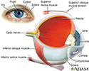
Eye - illustration
The eye is the organ of sight, a nearly spherical hollow globe filled with fluids (humors). The outer layer (sclera, or white of the eye, and cornea) is fibrous and protective. The middle layer (choroid, ciliary body and the iris) is vascular. The innermost layer (retina) is sensory nerve tissue that is light sensitive. The fluids in the eye are divided by the lens into the vitreous humor (behind the lens) and the aqueous humor (in front of the lens). The lens itself is flexible and suspended by ligaments which allow it to change shape to focus light on the retina, which is composed of sensory neurons.
Eye
illustration
-
Cataract - close-up of the eye - illustration
This photograph shows a cloudy white lens (cataract) as seen through the pupil. Cataracts are a leading cause of decreased vision in older adults, but children may have congenital cataracts. With surgery, the cataract can be removed, a new lens implanted, and the person can usually return home the same day.
Cataract - close-up of the eye
illustration
-
Rubella Syndrome - illustration
Rubella syndrome, or congenital rubella, is a group of physical abnormalities that have developed in an infant as a result of maternal infection and subsequent fetal infection with rubella virus. It is characterized by rash at birth, low birth weight, small head size, heart abnormalities, visual problems and bulging fontanelle.
Rubella Syndrome
illustration
-
Cataract - illustration
The lens of an eye is normally clear. If the lens becomes cloudy (opacified) it is called a cataract.
Cataract
illustration
-
Eye - illustration
The eye is the organ of sight, a nearly spherical hollow globe filled with fluids (humors). The outer layer (sclera, or white of the eye, and cornea) is fibrous and protective. The middle layer (choroid, ciliary body and the iris) is vascular. The innermost layer (retina) is sensory nerve tissue that is light sensitive. The fluids in the eye are divided by the lens into the vitreous humor (behind the lens) and the aqueous humor (in front of the lens). The lens itself is flexible and suspended by ligaments which allow it to change shape to focus light on the retina, which is composed of sensory neurons.
Eye
illustration
-
Cataract - close-up of the eye - illustration
This photograph shows a cloudy white lens (cataract) as seen through the pupil. Cataracts are a leading cause of decreased vision in older adults, but children may have congenital cataracts. With surgery, the cataract can be removed, a new lens implanted, and the person can usually return home the same day.
Cataract - close-up of the eye
illustration
-
Rubella Syndrome - illustration
Rubella syndrome, or congenital rubella, is a group of physical abnormalities that have developed in an infant as a result of maternal infection and subsequent fetal infection with rubella virus. It is characterized by rash at birth, low birth weight, small head size, heart abnormalities, visual problems and bulging fontanelle.
Rubella Syndrome
illustration
-
Cataract - illustration
The lens of an eye is normally clear. If the lens becomes cloudy (opacified) it is called a cataract.
Cataract
illustration
Review Date: 8/4/2023
Reviewed By: Franklin W. Lusby, MD, Ophthalmologist, Lusby Vision Institute, La Jolla, CA. Also reviewed by David C. Dugdale, MD, Medical Director, Brenda Conaway, Editorial Director, and the A.D.A.M. Editorial team.


