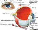
Ciliary body
The ciliary body is a circular structure that is an extension of the iris, the colored part of the eye. The ciliary body produces the fluid in the eye called aqueous humor. It also contains the ciliary muscle, which changes the shape of the lens when your eyes focus on a near object. This process is called accommodation.
Iris
The iris is the colored part of the eye. It is located between the cornea and lens. The round, central opening of the iris is called the pupil. Ve...

References
Evans M. Anatomy of the uvea. In: Yanoff M, Duker JS, eds. Ophthalmology. 6th ed. Philadelphia, PA: Elsevier; 2023:chap 7.1.
Standring S. Eye. In: Standring S, ed. Gray's Anatomy: The Anatomical Basis of Clinical Practice. 42nd ed. Philadelphia, PA: Elsevier; 2021:chap 45.
-
Eye - illustration
The eye is the organ of sight, a nearly spherical hollow globe filled with fluids (humors). The outer layer (sclera, or white of the eye, and cornea) is fibrous and protective. The middle layer (choroid, ciliary body and the iris) is vascular. The innermost layer (retina) is sensory nerve tissue that is light sensitive. The fluids in the eye are divided by the lens into the vitreous humor (behind the lens) and the aqueous humor (in front of the lens). The lens itself is flexible and suspended by ligaments which allow it to change shape to focus light on the retina, which is composed of sensory neurons.
Eye
illustration
-
Ciliary body - illustration
The ciliary body is a ring of tissue that encircles the lens. The ciliary body contains smooth muscle fibers called ciliary muscles that help to control the shape of the lens. Towards the posterior surface of the lens there are ciliary processes which contain capillaries. The capillaries secrete the fluid (vitreous humor) into the anterior segment of the eyeball.
Ciliary body
illustration
-
Eye - illustration
The eye is the organ of sight, a nearly spherical hollow globe filled with fluids (humors). The outer layer (sclera, or white of the eye, and cornea) is fibrous and protective. The middle layer (choroid, ciliary body and the iris) is vascular. The innermost layer (retina) is sensory nerve tissue that is light sensitive. The fluids in the eye are divided by the lens into the vitreous humor (behind the lens) and the aqueous humor (in front of the lens). The lens itself is flexible and suspended by ligaments which allow it to change shape to focus light on the retina, which is composed of sensory neurons.
Eye
illustration
-
Ciliary body - illustration
The ciliary body is a ring of tissue that encircles the lens. The ciliary body contains smooth muscle fibers called ciliary muscles that help to control the shape of the lens. Towards the posterior surface of the lens there are ciliary processes which contain capillaries. The capillaries secrete the fluid (vitreous humor) into the anterior segment of the eyeball.
Ciliary body
illustration
Review Date: 8/4/2023
Reviewed By: Franklin W. Lusby, MD, Ophthalmologist, Lusby Vision Institute, La Jolla, CA. Also reviewed by David C. Dugdale, MD, Medical Director, Brenda Conaway, Editorial Director, and the A.D.A.M. Editorial team.


