Arteriogram
Angiogram; AngiographyAn arteriogram is an imaging test that uses x-rays and a special dye to see inside the arteries. It can be used to view arteries in the heart, brain, kidney, and other parts of the body.
Related tests include:
-
Aortic angiography (chest or abdomen)
Aortic angiography
Aortic angiography is a procedure that uses a special dye and x-rays to see how blood flows through the aorta. The aorta is the large artery that ca...
 ImageRead Article Now Book Mark Article
 -
Cerebral angiography (brain)
Cerebral angiography
Cerebral angiography is a procedure that uses a special dye (contrast material) and x-rays to see how blood flows through the brain.
 ImageRead Article Now Book Mark Article
 -
Coronary angiography (heart)
Coronary angiography
Coronary angiography is a procedure that uses a special dye (contrast material) and x-rays to see how blood flows through the arteries in your heart....
 ImageRead Article Now Book Mark Article
 -
Extremity angiography (legs or arms)
Extremity angiography
Extremity angiography is a test used to see the arteries in the hands, arms, feet, or legs. It is also called peripheral angiography. Angiography u...
Read Article Now Book Mark Article -
Pulmonary angiography (lungs)
Pulmonary angiography
Pulmonary angiography is a test to see how blood flows through the lung. Angiography is an imaging test that uses x-rays and a special dye to see th...
 ImageRead Article Now Book Mark Article
 -
Renal arteriography (kidneys)
Renal arteriography
Renal arteriography is a special x-ray of the arteries of the kidneys.
 ImageRead Article Now Book Mark Article
 -
Mesenteric angiography (colon or small bowel)
Mesenteric angiography
Mesenteric angiography is a test used to look at the blood vessels that supply the stomach, small and large intestines, and other internal organs. An...
 ImageRead Article Now Book Mark Article
 - Pelvic angiography (pelvis)
- Hepatic angiography (liver)
How the Test is Performed
The test is done in a medical facility designed to perform this test. You will lie on an x-ray table. Local anesthetic is used to numb the area where the dye is injected. Most of the time, an artery in the groin will be used. In some cases, an artery in your wrist may be used.
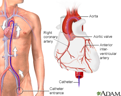
Next, a flexible tube called a catheter (which is the width of the tip of a pen) is inserted into the artery in the groin and moved through the artery until it reaches the intended area of the body. The exact procedure depends on the part of the body being examined.
You will not feel the catheter inside of you.
You may ask for a calming medicine (sedative) if you are anxious about the test.
For most tests:
- A dye (contrast) is injected into an artery.
- X-rays are taken to see how the dye flows through your bloodstream.
How to Prepare for the Test
How you should prepare depends on the part of the body being examined. Your health care provider may tell you to stop taking certain medicines that could affect the test, or blood thinning medicines. DO NOT stop taking any medicine without first talking to your provider. In most cases, you may not be able to eat or drink anything for a few hours before the test.
How the Test will Feel
You may have some discomfort from a needle stick. The needle stick will usually be in the groin or wrist. You may feel symptoms such as flushing in the face or other parts of the body when the dye is injected. The exact symptoms will depend on the part of the body being examined.
If you had an injection in your groin area, you will most often be asked to lie flat on your back for a few hours after the test. This is to help avoid bleeding. Lying flat may be uncomfortable for some people.
Why the Test is Performed
An arteriogram is done to see how blood moves through the arteries. It is also used to check for blocked or damaged arteries. It can be used to visualize tumors or find a source of bleeding. Usually, an arteriogram is performed at the same time as a treatment. If no treatment is planned, in many areas of the body it has been replaced with CT or MR arteriography.
References
Azarbal AF, Mclafferty RB. Arteriography. In: Sidawy AN, Perler BA, eds. Rutherford's Vascular Surgery and Endovascular Therapy. 10th ed. Philadelphia, PA: Elsevier; 2023:chap 27.
DeCarlo TE, Olson JL, Mandava N. Camera-based ancillary retinal testing: autofluorescence, fluorescein, and indocyanine green angiography. In: Yanoff M, Duker JS, eds. Ophthalmology. 6th ed. Philadelphia, PA: Elsevier; 2023:chap 6.4.
Harisinghani MG. Chen JW, Weissleder R. Vascular imaging. In: Harisinghani MG. Chen JW, Weissleder R, eds. Primer of Diagnostic Imaging. 6th ed. Philadelphia, PA: Elsevier; 2019:chap 8.
Mondschein JI, Solomon JA. Peripheral arterial disease diagnosis and intervention. In: Torigian DA, Ramchandani P, eds. Radiology Secrets Plus. 4th ed. Philadelphia, PA: Elsevier; 2017:chap 70.
-
Cardiac arteriogram - illustration
An arteriogram is the injection of contrast material or dye into one or more arteries to make them visible on an x-ray. The blood flow through the area can be evaluated with fluoroscopy (i.e., continuous X-rays that allow one to see the contrast material in movement).
Cardiac arteriogram
illustration
Review Date: 1/29/2024
Reviewed By: Jason Levy, MD, FSIR, Northside Radiology Associates, Atlanta, GA. Also reviewed by David C. Dugdale, MD, Medical Director, Brenda Conaway, Editorial Director, and the A.D.A.M. Editorial team.


