X-ray - skeleton
Skeletal surveyA skeletal x-ray is an imaging test used to look at your bones. It is used to detect fractures, tumors, or conditions that cause wearing away (degeneration) of the bone.
x-ray
X-rays are a type of electromagnetic radiation, just like visible light. An x-ray machine sends individual x-ray waves through the body. The images...

Fractures
If more pressure is put on a bone than it can stand, it will split or break. A break of any size is called a fracture. If the broken bone punctures...

How the Test is Performed
The test is done in a hospital radiology department or in your health care provider's office by an x-ray technologist.
You will lie on a table or stand in front of the x-ray machine, depending on the bone that is injured. You may be asked to change position so that different x-ray views can be taken.
The x-rays pass through your body. A computer or special film records the images.
Structures that are dense (such as bone) will block most of the x-ray particles. These areas will appear white. Metal and contrast media (special dye used to highlight areas of the body) will also appear white. Structures containing air will be black. Muscle, fat, and fluid will appear as shades of gray.
How to Prepare for the Test
Tell your provider if you are pregnant. You must remove all jewelry before the x-ray.
How the Test will Feel
The x-rays are painless. Changing positions and moving the injured area for different x-ray views may be uncomfortable. If the whole skeleton is being imaged, the test most often takes 1 hour or more.
Why the Test is Performed
This test is used to look for:
- Fractures or broken bone
- Cancer that has spread to other areas of the body
-
Osteomyelitis (inflammation of the bone caused by an infection)
Osteomyelitis
Osteomyelitis is a bone infection. It is caused by bacteria or other germs.
 ImageRead Article Now Book Mark Article
 - Bone damage due to trauma (such as an auto accident) or degenerative conditions
- Abnormalities in the soft tissue around the bone
What Abnormal Results Mean
Abnormal findings include:
- Fractures
-
Bone tumors
Bone tumors
A bone tumor is an abnormal growth of cells within a bone. A bone tumor may be cancerous (malignant) or noncancerous (benign).
 ImageRead Article Now Book Mark Article
 - Degenerative bone conditions
- Osteomyelitis
Risks
There is low radiation exposure. X-rays machines are set to provide the smallest amount of radiation exposure needed to produce the image. Most experts feel that the risk is low compared with the benefits.
Children and the fetuses of pregnant women are more sensitive to the risks of the x-ray. A protective shield may be worn over areas not being scanned.
References
Contreras F, Perez J, Jose J. Imaging overview. In: Miller MD, Thompson SR, eds. DeLee, Drez, & Miller's Orthopaedic Sports Medicine. 5th ed. Philadelphia, PA: Elsevier; 2020:chap 7.
Kapoor G, Toms AP. Current status of imaging of the musculoskeletal system. In: Adam A, Dixon AK, Gillard JH, Schaefer-Prokop CM, eds. Grainger & Allison's Diagnostic Radiology. 7th ed. Philadelphia, PA: Elsevier; 2021:chap 38.
-
X-ray - illustration
X-rays are a form of ionizing radiation that can penetrate the body to form an image on film. Structures that are dense (such as bone) will appear white, air will be black, and other structures will be shades of gray depending on density. X-rays can provide information about obstructions, tumors, and other diseases, especially when coupled with the use of barium and air contrast within the bowel.
X-ray
illustration
-
Skeleton - illustration
The skeleton consists of groups of bones which protect and move the body.
Skeleton
illustration
-
Skeletal spine - illustration
The spine is divided into several sections. The cervical vertebrae make up the neck. The thoracic vertebrae comprise the chest section and have ribs attached. The lumbar vertebrae are the remaining vertebrae below the last thoracic bone and the top of the sacrum. The sacral vertebrae are caged within the bones of the pelvis, and the coccyx represents the terminal vertebrae or vestigial tail.
Skeletal spine
illustration
-
Hand X-ray - illustration
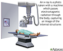
An x-ray is a photo taken with a machine which passes electromagnetic radiation through the body, capturing an image of the internal structures.
Hand X-ray
illustration
-
Skeleton (posterior view) - illustration
The skeleton is made up of 206 bones in the adult and contributes to the form and shape of the body. The skeleton has several important functions for the body. The bones of the skeleton provide support for the soft tissues. For example, the rib cage supports the thoracic wall. Most muscles of the body are attached to bones which act as levers to allow movement of body parts. The bones of the skeleton also serve as a reservoir for minerals, such as calcium and phosphate. Finally, most of the blood cell formation takes places within the marrow of certain bones.
Skeleton (posterior view)
illustration
-
The skeleton (lateral view) - illustration
The skeleton is made up of 206 bones in the adult and contributes to the form and shape of the body. The skeleton has several important functions for the body. The bones of the skeleton provide support for the soft tissues. For example, the rib cage supports the thoracic wall. Most muscles of the body are attached to bones which act as levers to allow movement of body parts. The bones of the skeleton also serve as a reservoir for minerals, such as calcium and phosphate. Finally, most of the blood cell formation takes places within the marrow of certain bones.
The skeleton (lateral view)
illustration
-
X-ray - illustration
X-rays are a form of ionizing radiation that can penetrate the body to form an image on film. Structures that are dense (such as bone) will appear white, air will be black, and other structures will be shades of gray depending on density. X-rays can provide information about obstructions, tumors, and other diseases, especially when coupled with the use of barium and air contrast within the bowel.
X-ray
illustration
-
Skeleton - illustration
The skeleton consists of groups of bones which protect and move the body.
Skeleton
illustration
-
Skeletal spine - illustration
The spine is divided into several sections. The cervical vertebrae make up the neck. The thoracic vertebrae comprise the chest section and have ribs attached. The lumbar vertebrae are the remaining vertebrae below the last thoracic bone and the top of the sacrum. The sacral vertebrae are caged within the bones of the pelvis, and the coccyx represents the terminal vertebrae or vestigial tail.
Skeletal spine
illustration
-
Hand X-ray - illustration
An x-ray is a photo taken with a machine which passes electromagnetic radiation through the body, capturing an image of the internal structures.
Hand X-ray
illustration
-
Skeleton (posterior view) - illustration
The skeleton is made up of 206 bones in the adult and contributes to the form and shape of the body. The skeleton has several important functions for the body. The bones of the skeleton provide support for the soft tissues. For example, the rib cage supports the thoracic wall. Most muscles of the body are attached to bones which act as levers to allow movement of body parts. The bones of the skeleton also serve as a reservoir for minerals, such as calcium and phosphate. Finally, most of the blood cell formation takes places within the marrow of certain bones.
Skeleton (posterior view)
illustration
-
The skeleton (lateral view) - illustration
The skeleton is made up of 206 bones in the adult and contributes to the form and shape of the body. The skeleton has several important functions for the body. The bones of the skeleton provide support for the soft tissues. For example, the rib cage supports the thoracic wall. Most muscles of the body are attached to bones which act as levers to allow movement of body parts. The bones of the skeleton also serve as a reservoir for minerals, such as calcium and phosphate. Finally, most of the blood cell formation takes places within the marrow of certain bones.
The skeleton (lateral view)
illustration
Review Date: 4/1/2025
Reviewed By: Linda J. Vorvick, MD, Clinical Professor Emeritus, Department of Family Medicine, UW Medicine, School of Medicine, University of Washington, Seattle, WA. Also reviewed by David C. Dugdale, MD, Medical Director, Brenda Conaway, Editorial Director, and the A.D.A.M. Editorial team.


