Calcium - ionized
Free calcium; Ionized calciumIonized calcium is calcium in your blood that is not attached to proteins. It is also called free calcium.
All cells need calcium in order to work. Calcium helps build strong bones and teeth. It is important for heart function. It also helps with muscle contraction, nerve signaling, and blood clotting.
This article discusses the test used to measure the amount of ionized calcium in blood.
How the Test is Performed
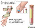
A blood sample is needed. Most of the time blood is drawn from a vein located on the inside of the elbow or the back of the hand.
Drawn from a vein
Venipuncture is the collection of blood from a vein. It is most often done for laboratory testing.

How to Prepare for the Test
Many medicines can interfere with blood test results.
- Your health care provider will tell you if you need to stop taking any medicines before you have this test.
- Do not stop or change your medicines without talking to your provider first.
Why the Test is Performed
Your provider may order this test if you have signs of bone, kidney, liver or parathyroid disease. The test may also be done to monitor the progress and treatment of these diseases.
Most of the time, blood tests measure your total calcium level. This looks at both ionized calcium and calcium attached to proteins. You may need to have a separate ionized calcium test if you have factors that increase or decrease total calcium levels. These may include abnormal blood levels of albumin or immunoglobulins.
Normal Results
Results generally fall in these ranges:
- Children: 4.8 to 5.3 milligrams per deciliter (mg/dL) or 1.20 to 1.32 millimoles per liter (millimol/L)
- Adults: 4.8 to 5.6 mg/dL or 1.20 to 1.40 millimol/L
Normal value ranges may vary slightly among different labs. Talk to your provider about the meaning of your specific test results.
The examples above show the common measurements for results for these tests. Some labs use different measurements or may test different specimens.
What Abnormal Results Mean
Higher-than-normal levels of ionized calcium may be due to:
- Decreased levels of calcium in the urine from an unknown cause (also called hypocalciuria)
-
Hyperparathyroidism
Hyperparathyroidism
Hyperparathyroidism is a condition in which 1 or more of the parathyroid glands in your neck produce too much parathyroid hormone (PTH).
 ImageRead Article Now Book Mark Article
 -
Hyperthyroidism
Hyperthyroidism
Hyperthyroidism is a condition in which the thyroid gland makes too much thyroid hormone. The condition is often called overactive thyroid.
 ImageRead Article Now Book Mark Article
 -
Milk-alkali syndrome
Milk-alkali syndrome
Milk-alkali syndrome is a condition in which there is a high level of calcium in the body (hypercalcemia). This causes a shift in the body's acid/ba...
Read Article Now Book Mark Article -
Multiple myeloma
Multiple myeloma
Multiple myeloma is a blood cancer that starts from a type of white blood cell in the bone marrow called plasma cells. Bone marrow is the soft, spon...
 ImageRead Article Now Book Mark Article
 -
Paget disease
Paget disease
Paget disease is a disorder that involves abnormal bone destruction and regrowth. This results in deformity of the affected bones.
 ImageRead Article Now Book Mark Article
 -
Sarcoidosis
Sarcoidosis
Sarcoidosis is a disease in which inflammation occurs in the lymph nodes, lungs, liver, eyes, skin, and other tissues.
 ImageRead Article Now Book Mark Article
 - Thiazide diuretics
- Thrombocytosis (high platelet count)
-
Tumors
Tumors
A tumor is an abnormal growth of body tissue. Tumors can be cancerous (malignant) or noncancerous (benign).
Read Article Now Book Mark Article -
Vitamin A excess
Vitamin A
Vitamin A is a fat-soluble vitamin that is stored in the liver. There are two types of vitamin A that are found in the diet. Preformed vitamin A is f...
 ImageRead Article Now Book Mark Article
 -
Vitamin D excess
Vitamin D
Vitamin D is a fat-soluble vitamin. Fat-soluble vitamins are stored in the body's fatty tissue and liver.
 ImageRead Article Now Book Mark Article
Lower-than-normal levels may be due to (or caused by):
-
Hypoparathyroidism
Hypoparathyroidism
Hypoparathyroidism is a disorder in which the parathyroid glands in the neck do not produce enough parathyroid hormone (PTH).
 ImageRead Article Now Book Mark Article
 -
Malabsorption
Malabsorption
Malabsorption involves problems with the body's ability to take in (absorb) nutrients from food.
 ImageRead Article Now Book Mark Article
 -
Osteomalacia
Osteomalacia
Osteomalacia is softening of the bones. It most often occurs because of a problem that leads to vitamin D deficiency, which helps your body absorb c...
 ImageRead Article Now Book Mark Article
 - Pancreatitis
-
Renal failure
Renal failure
Acute kidney failure is the rapid (less than 2 days) loss of your kidneys' ability to remove waste and help balance fluids and electrolytes in your b...
 ImageRead Article Now Book Mark Article
 -
Rickets
Rickets
Rickets is a disorder that occurs in children before bone growth is complete. It is caused by a lack of vitamin D, calcium, or phosphate. It leads ...
 ImageRead Article Now Book Mark Article
 - Vitamin D deficiency
References
Bilezikian JP, Walker MD, Binkley N, Goltzman D, Mannstadt M. Hormones and disorders of mineral metabolism. In: Melmed S, Auchus, RJ, Goldfine AB, Rosen CJ, Kopp PA, eds. Williams Textbook of Endocrinology. 15th ed. Philadelphia, PA: Elsevier; 2025:chap 27.
Klemm KM, Klein MJ, Zhang Y. Biochemical markers of bone metabolism. In: McPherson RA, Pincus MR, eds. Henry's Clinical Diagnosis and Management by Laboratory Methods. 24th ed. Philadelphia, PA: Elsevier; 2022:chap 16.
Thakker RV. The parathyroid glands, hypercalcemia, and hypocalcemia. In: Goldman L, Cooney KA, eds. Goldman-Cecil Medicine. 27th ed. Philadelphia, PA: Elsevier; 2024:chap 227.
-
Blood test - illustration
Blood is drawn from a vein (venipuncture), usually from the inside of the elbow or the back of the hand. A needle is inserted into the vein, and the blood is collected in an air-tight vial or a syringe. Preparation may vary depending on the specific test.
Blood test
illustration
Review Date: 5/19/2025
Reviewed By: Jacob Berman, MD, MPH, Clinical Assistant Professor of Medicine, Division of General Internal Medicine, University of Washington School of Medicine, Seattle, WA. Also reviewed by David C. Dugdale, MD, Medical Director, Brenda Conaway, Editorial Director, and the A.D.A.M. Editorial team.


