Lyme disease blood test
Lyme disease serology; ELISA for Lyme disease; Western blot for Lyme diseaseThe Lyme disease blood test looks for antibodies in the blood to the bacteria that causes Lyme disease. The test is used to help diagnose Lyme disease.
Lyme disease
Lyme disease is a bacterial infection that is spread through the bite of one of several types of ticks.

How the Test is Performed
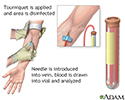
A blood sample is needed.
Blood sample
Venipuncture is the collection of blood from a vein. It is most often done for laboratory testing.

A laboratory specialist looks for Lyme disease antibodies in the blood sample using the ELISA test. If the ELISA test is positive, it must be confirmed with another test called the Western blot test.
ELISA test
ELISA stands for enzyme-linked immunoassay. It is a commonly used laboratory test to detect antibodies in the blood. An antibody is a protein produ...

How to Prepare for the Test
You do not need to take special steps to prepare for this test.
How the Test will Feel
When the needle is inserted to draw blood, some people feel moderate pain. Others feel only a prick or stinging. Afterward, there may be some throbbing or a slight bruise. This soon goes away.
Why the Test is Performed
The test is done to check for the diagnosis of Lyme disease.
Normal Results
A negative test result is normal. This means none or few antibodies to Lyme disease were seen in your blood sample. If the ELISA test is negative, usually no other testing is needed.
Normal value ranges may vary slightly among different laboratories. Some labs use different measurements or test different samples. Talk to your health care provider about the meaning of your specific test results.
What Abnormal Results Mean
A positive ELISA result is abnormal. This means antibodies were seen in your blood sample. But, this does not confirm a diagnosis of Lyme disease. A positive ELISA result must be followed up with a Western blot test. Only a positive Western blot test can confirm the diagnosis of Lyme disease.
For many people, the ELISA test remains positive, even after they have been treated for Lyme disease and no longer have symptoms.
A positive ELISA test may also occur with certain diseases not related to Lyme disease, such as rheumatoid arthritis.
Risks
There is little risk involved with having your blood taken. Veins and arteries vary in size from one person to another and from one side of the body to the other. Taking blood from some people may be more difficult than from others.
Other risks associated with having blood drawn are slight but may include:
- Fainting or feeling lightheaded
- Multiple punctures to locate veins
-
Hematoma (blood buildup under the skin)
Hematoma
A bruise is an area of skin discoloration. A bruise occurs when small blood vessels break and leak their contents into the soft tissue beneath the s...
 ImageRead Article Now Book Mark Article
 - Excessive bleeding
- Infection (a slight risk any time the skin is broken)
References
Nikolic D. Spirochete infections. In: McPherson RA, Pincus MR, eds. Henry's Clinical Diagnosis and Management by Laboratory Methods. 24th ed. Philadelphia, PA: Elsevier; 2022:chap 61.
Steere AC. Lyme disease (Lyme borreliosis) due to Borrelia burgdorferi. In: Bennett JE, Dolin R, Blaser MJ, eds. Mandell, Douglas, and Bennett's Principles and Practice of Infectious Diseases. 9th ed. Philadelphia, PA: Elsevier; 2020:chap 241.
-
Blood test - illustration
Blood is drawn from a vein (venipuncture), usually from the inside of the elbow or the back of the hand. A needle is inserted into the vein, and the blood is collected in an air-tight vial or a syringe. Preparation may vary depending on the specific test.
Blood test
illustration
-
Lyme disease organism, Borrelia burgdorferi - illustration
Borrelia burgdorferi is a spirochete bacteria that causes Lyme disease. It is similar in shape to the spirochetes that cause other diseases, such as relapsing fever and syphilis. (Image courtesy of the Centers for Disease Control and Prevention.)
Lyme disease organism, Borrelia burgdorferi
illustration
-
Deer ticks - illustration
Diseases are often carried by ticks, including Rocky Mountain Spotted Fever, Colorado Tick Fever, Lyme disease, and tularemia. Less common or less frequent diseases include typhus, Q-fever, relapsing fever, viral encephalitis, hemorrhagic fever, and babesiosis.
Deer ticks
illustration
-
Ticks - illustration
There are many species of ticks. Of these, a large proportion are capable of carrying disease. Diseases carried by ticks include Lyme disease, Ehrlichiosis, Rocky Mountain Spotted Fever, Colorado Tick Fever, tularemia, typhus, hemorrhagic fever, and viral encephalitis. (Image courtesy of the Centers for Disease Control and Prevention.)
Ticks
illustration
-
Lyme disease - Borrelia burgdorferi organism - illustration
Lyme disease is caused when a person is bitten by a tick infected with bacteria called Borrelia burgdorferi. These bacteria are known as spirochetes because of their long, corkscrew shape. This photograph shows the typical corkscrew appearance of a spirochete. Other types of spirochete bacteria cause diseases such as syphilis and leptospirosis. After a person is bitten by an infected tick, the bacteria spread from the skin through the bloodstream to other organs. The skin, nervous system, joints, heart, and eyes are most often affected. (Image courtesy of the Centers for Disease Control and Prevention.)
Lyme disease - Borrelia burgdorferi organism
illustration
-
Tick imbedded in the skin - illustration
This is a close-up photograph of a tick embedded in the skin. Ticks are important because they can carry diseases such as Rocky Mountain spotted fever, tularemia, Colorado tick fever, Lyme disease, and others.
Tick imbedded in the skin
illustration
-
Antibodies - illustration
Antigens are large molecules (usually proteins) on the surface of cells, viruses, fungi, bacteria, and some non-living substances such as toxins, chemicals, drugs, and foreign particles. The immune system recognizes antigens and produces antibodies that destroy substances containing antigens.
Antibodies
illustration
-
Blood test - illustration
Blood is drawn from a vein (venipuncture), usually from the inside of the elbow or the back of the hand. A needle is inserted into the vein, and the blood is collected in an air-tight vial or a syringe. Preparation may vary depending on the specific test.
Blood test
illustration
-
Lyme disease organism, Borrelia burgdorferi - illustration
Borrelia burgdorferi is a spirochete bacteria that causes Lyme disease. It is similar in shape to the spirochetes that cause other diseases, such as relapsing fever and syphilis. (Image courtesy of the Centers for Disease Control and Prevention.)
Lyme disease organism, Borrelia burgdorferi
illustration
-
Deer ticks - illustration
Diseases are often carried by ticks, including Rocky Mountain Spotted Fever, Colorado Tick Fever, Lyme disease, and tularemia. Less common or less frequent diseases include typhus, Q-fever, relapsing fever, viral encephalitis, hemorrhagic fever, and babesiosis.
Deer ticks
illustration
-
Ticks - illustration
There are many species of ticks. Of these, a large proportion are capable of carrying disease. Diseases carried by ticks include Lyme disease, Ehrlichiosis, Rocky Mountain Spotted Fever, Colorado Tick Fever, tularemia, typhus, hemorrhagic fever, and viral encephalitis. (Image courtesy of the Centers for Disease Control and Prevention.)
Ticks
illustration
-
Lyme disease - Borrelia burgdorferi organism - illustration
Lyme disease is caused when a person is bitten by a tick infected with bacteria called Borrelia burgdorferi. These bacteria are known as spirochetes because of their long, corkscrew shape. This photograph shows the typical corkscrew appearance of a spirochete. Other types of spirochete bacteria cause diseases such as syphilis and leptospirosis. After a person is bitten by an infected tick, the bacteria spread from the skin through the bloodstream to other organs. The skin, nervous system, joints, heart, and eyes are most often affected. (Image courtesy of the Centers for Disease Control and Prevention.)
Lyme disease - Borrelia burgdorferi organism
illustration
-
Tick imbedded in the skin - illustration
This is a close-up photograph of a tick embedded in the skin. Ticks are important because they can carry diseases such as Rocky Mountain spotted fever, tularemia, Colorado tick fever, Lyme disease, and others.
Tick imbedded in the skin
illustration
-
Antibodies - illustration
Antigens are large molecules (usually proteins) on the surface of cells, viruses, fungi, bacteria, and some non-living substances such as toxins, chemicals, drugs, and foreign particles. The immune system recognizes antigens and produces antibodies that destroy substances containing antigens.
Antibodies
illustration
Review Date: 12/31/2023
Reviewed By: Jatin M. Vyas, MD, PhD, Roy and Diana Vagelos Professor in Medicine, Columbia University Vagelos College of Physicians and Surgeons, Division of Infectious Diseases, Department of Medicine, New York, NY. Also reviewed by David C. Dugdale, MD, Medical Director, Brenda Conaway, Editorial Director, and the A.D.A.M. Editorial team.


