Acetylcholine receptor antibody
Acetylcholine receptor antibody is a protein found in the blood of many people with myasthenia gravis. The antibody affects the part of the muscle that receives signals from nerves.
Myasthenia gravis
Myasthenia gravis is a neuromuscular disorder. Neuromuscular disorders involve the muscles and the nerves that control them.

Antibody
An antibody is a protein produced by the body's immune system when it detects harmful substances, called antigens. Examples of antigens include micr...

This article discusses the blood test for acetylcholine receptor antibody.
How the Test is Performed
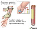
A blood sample is needed. Most of the time, blood is drawn from a vein located on the inside of the elbow or the back of the hand.
Drawn from a vein
Venipuncture is the collection of blood from a vein. It is most often done for laboratory testing.

How to Prepare for the Test
Most of the time, you do not need to take special steps before this test.
How the Test will Feel
You may feel slight pain or a sting when the needle is inserted. You may also feel some throbbing at the site after the blood is drawn.
Why the Test is Performed
This test is used to help diagnose myasthenia gravis.
Normal Results
Normally, there is no acetylcholine receptor antibody (or less than 0.02 nmol/L) in the bloodstream.
Note: Normal value ranges may vary slightly among different labs. Talk to your health care provider about the meaning of your specific test results.
What Abnormal Results Mean
An abnormal result means acetylcholine receptor antibody has been found in your blood. It confirms the diagnosis of myasthenia gravis in people who have symptoms of myasthenia gravis. Nearly one half of people with myasthenia gravis that is limited to their eye muscles (ocular myasthenia gravis) have this antibody in their blood. This antibody can also be present in the blood of people who have a thymus gland tumor (thymoma), with or without myasthenia gravis.
However, the lack of this antibody does not rule out myasthenia gravis. About 1 in 5 people with myasthenia gravis do not have signs of this antibody in their blood. Your provider may also consider testing you for the muscle specific kinase (MuSK) or other antibodies.
References
Guptill JT, Sanders DB. Disorders of neuromuscular transmission. In: Jankovic J, Mazziotta JC, Pomeroy SL, Newman NJ, eds. Bradley and Daroff's Neurology in Clinical Practice. 8th ed. Philadelphia, PA: Elsevier; 2022:chap 108.
Kaminski HJ. Disorders of neuromuscular transmission. In: Goldman, Cooney KA, eds. Goldman-Cecil Medicine. 27th ed. Philadelphia, PA: Elsevier; 2024:chap 390.
-
Blood test - illustration
Blood is drawn from a vein (venipuncture), usually from the inside of the elbow or the back of the hand. A needle is inserted into the vein, and the blood is collected in an air-tight vial or a syringe. Preparation may vary depending on the specific test.
Blood test
illustration
-
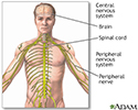
Central nervous system and peripheral nervous system - illustration
The central nervous system comprises the brain and spinal cord. The peripheral nervous system includes nerves outside the brain and spinal cord.
Central nervous system and peripheral nervous system
illustration
-
Ptosis - drooping of the eyelid - illustration
Drooping of the eyelid is called ptosis. Ptosis may result from damage to the nerve that controls the muscles of the eyelid, problems with the muscle strength (as in myasthenia gravis), or from swelling of the lid.
Ptosis - drooping of the eyelid
illustration
-
Blood test - illustration
Blood is drawn from a vein (venipuncture), usually from the inside of the elbow or the back of the hand. A needle is inserted into the vein, and the blood is collected in an air-tight vial or a syringe. Preparation may vary depending on the specific test.
Blood test
illustration
-
Central nervous system and peripheral nervous system - illustration
The central nervous system comprises the brain and spinal cord. The peripheral nervous system includes nerves outside the brain and spinal cord.
Central nervous system and peripheral nervous system
illustration
-
Ptosis - drooping of the eyelid - illustration
Drooping of the eyelid is called ptosis. Ptosis may result from damage to the nerve that controls the muscles of the eyelid, problems with the muscle strength (as in myasthenia gravis), or from swelling of the lid.
Ptosis - drooping of the eyelid
illustration
Review Date: 4/16/2025
Reviewed By: Joseph V. Campellone, MD, Department of Neurology, Cooper Medical School at Rowan University, Camden, NJ. Review provided by VeriMed Healthcare Network. Also reviewed by David C. Dugdale, MD, Medical Director, Brenda Conaway, Editorial Director, and the A.D.A.M. Editorial team.


