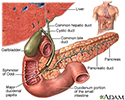
Gallbladder radionuclide scan
Radionuclide - gallbladder; Gallbladder scan; Biliary scan; Cholescintigraphy; HIDA; Hepatobiliary nuclear imaging scanGallbladder radionuclide scan is a test that uses radioactive material to check gallbladder function. It is also used to look for bile duct blockage or leak.
How the Test is Performed
The health care provider will inject a radioactive chemical called a gamma-emitting tracer into a vein. This material collects mostly in the liver. It will then flow with bile into the gallbladder and then through the common bile duct to the duodenum or small intestine.
For the test:
- You lie face up on a table under a scanner called a gamma camera. The scanner detects the rays coming from the tracer. A computer displays images of where the tracer is found in the organs.
- Images are taken every 5 to 15 minutes. Most of the time, the test takes about 1 hour. At times, it can take up to 4 hours.
If the provider cannot see the gallbladder after certain amount of time, you may be given a small amount of morphine. This can help the radioactive material get into the gallbladder. The morphine may cause you to feel tired after the exam.
In some cases, you may be given a medicine during this test to see how well your gallbladder squeezes (contracts). The medicine may be injected into the vein. Otherwise, you may be asked to drink a high-density drink like Boost which will help your gallbladder contract.
How to Prepare for the Test
You need to eat something within a day of the test. However, you must stop eating or drinking 4 hours before the test starts.
How the Test will Feel
You will feel a sharp prick from the needle when the tracer is injected into the vein. The site may be sore after the injection. There is normally no pain during the scan.
Why the Test is Performed
This test is very good for detecting a sudden infection of the gallbladder or blockage of a bile duct. It is also helpful in determining whether there is a complication of a transplanted liver or a leak after the gallbladder has been surgically removed.
The test can also be used to detect long-term gallbladder problems.
What Abnormal Results Mean
Abnormal results may be due to:
- Abnormal anatomy of the bile system (biliary anomalies)
-
Bile duct obstruction
Bile duct obstruction
Bile duct obstruction is a blockage in the tubes that carry bile from the liver to the gallbladder and small intestine.
 ImageRead Article Now Book Mark Article
 - Bile leaks or abnormal ducts
-
Cancer of the hepatobiliary system
Cancer of the hepatobiliary system
Hepatocellular carcinoma is cancer that starts in the liver.
 ImageRead Article Now Book Mark Article
 - Gallbladder infection (cholecystitis)
Cholecystitis
Acute cholecystitis is sudden swelling and irritation of the gallbladder. It causes severe belly pain.
 ImageRead Article Now Book Mark Article
 -
Gallstones
Gallstones
Gallstones are hard deposits that form inside the gallbladder. These may be as small as a grain of sand or as large as a golf ball.
 ImageRead Article Now Book Mark Article
 - Infection of the gallbladder, ducts, or liver
-
Liver disease
Liver disease
The term "liver disease" applies to many conditions that stop the liver from working or prevent it from functioning well. Abdominal pain or swelling...
 ImageRead Article Now Book Mark Article
 -
Transplant complication (after liver transplant)
Transplant complication
Transplant rejection is a process in which a transplant recipient's immune system attacks the transplanted organ or tissue.
 ImageRead Article Now Book Mark Article
Liver transplant
Liver transplant is surgery to replace a diseased liver with a healthy liver.
 ImageRead Article Now Book Mark Article
Risks
There is a small risk to pregnant or nursing mothers. Unless it is absolutely necessary, the scan will be delayed until you are no longer pregnant or nursing.
The amount of radiation is small (less than that of a regular x-ray). It is almost all gone from the body within 1 or 2 days. Your risk from radiation may increase if you have a lot of scans.
Considerations
Most of the time, this test is done only if a person has abdominal pain that may be from gallbladder disease or gallstones. For this reason, some people may need urgent treatment based on the test results.
This test is combined with other imaging (such as CT or ultrasound). After the gallbladder scan, the person may be prepared for surgery, if needed.
CT
An abdominal CT scan is an imaging test that uses x-rays to create cross-sectional pictures of the belly area. CT stands for computed tomography....

Ultrasound
Abdominal ultrasound is a type of imaging test. It is used to look at organs in the abdomen, including the liver, gallbladder, pancreas, and kidneys...

References
Grajo JR. Imaging of the liver. In: Sahani DV, Samir AE, eds. Abdominal Imaging. 2nd ed. Philadelphia, PA: Elsevier; 2017:chap 35.
O'Malley JP, Ziessman HA, Thrall JH. Hepatic, biliary, and splenic scintigraphy. In: O'Malley JP, Ziessman HA, Thrall JH, eds. Nuclear Medicine and Molecular Imaging: The Requisites. 5th ed. Philadelphia, PA: Elsevier; 2021:chap 9.
Wang DQH, Afdhal NH. Gallstone disease. In: Feldman M, Friedman LS, Brandt LJ, eds. Sleisenger and Fordtran's Gastrointestinal and Liver Disease. 11th ed. Philadelphia, PA: Elsevier; 2021:chap 65.
-
Gallbladder - illustration
The gallbladder is a muscular sac located under the liver. It stores and concentrates the bile produced in the liver that is not immediately needed for digestion. Bile is released from the gallbladder into the small intestine in response to food. The pancreatic duct joins the common bile duct at the small intestine adding enzymes to aid in digestion.
Gallbladder
illustration
-
Gallbladder radionuclide scan - illustration
The gallbladder radionuclide scan is performed by injecting a tracer (radioactive chemical) into the bloodstream. A gamma camera is used to perform the scan. The camera will detect the gamma rays being emitted from the tracer, and the image of where the tracer is found in the organs is transmitted to a computer. This test is very good for detecting acute infection (cholecystitis) or blockage of a bile duct. It is also helpful in determining whether there is rejection of a transplanted liver.
Gallbladder radionuclide scan
illustration
-
Gallbladder - illustration
The gallbladder is a muscular sac located under the liver. It stores and concentrates the bile produced in the liver that is not immediately needed for digestion. Bile is released from the gallbladder into the small intestine in response to food. The pancreatic duct joins the common bile duct at the small intestine adding enzymes to aid in digestion.
Gallbladder
illustration
-
Gallbladder radionuclide scan - illustration
The gallbladder radionuclide scan is performed by injecting a tracer (radioactive chemical) into the bloodstream. A gamma camera is used to perform the scan. The camera will detect the gamma rays being emitted from the tracer, and the image of where the tracer is found in the organs is transmitted to a computer. This test is very good for detecting acute infection (cholecystitis) or blockage of a bile duct. It is also helpful in determining whether there is rejection of a transplanted liver.
Gallbladder radionuclide scan
illustration
Review Date: 1/1/2025
Reviewed By: Jason Levy, MD, FSIR, Northside Radiology Associates, Atlanta, GA. Also reviewed by David C. Dugdale, MD, Medical Director, Brenda Conaway, Editorial Director, and the A.D.A.M. Editorial team.


