Enteroscopy
Push enteroscopy; Double-balloon enteroscopy; Single-balloon enteroscopy; Spiral enteroscopy; Capsule enteroscopyEnteroscopy is a procedure used to examine the small intestine (small bowel).
How the Test is Performed
An enteroscope uses a long flexible tube with a balloon and a camera attached that can go deep down the inside of the small bowel.
During a single- or double-balloon enteroscopy, balloons attached to the endoscope can be inflated to allow the doctor to view a section of the small intestine. The enteroscope is usually inserted through the mouth for upper enteroscopy.
In a colonoscopy, a flexible tube is inserted through your rectum and colon. The tube can most often reach into the end part of the small intestine (ileum). Occasionally, an enteroscope is inserted through the rectum to reach further into the end of the small intestine.
Colonoscopy
A colonoscopy is an exam that views the inside of the colon (large intestine) and rectum, using a tool called a colonoscope. The colonoscope has a sm...

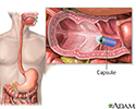
Capsule endoscopy is done with a disposable capsule that you swallow. This transmits pictures to a recorder and is uploaded to a computer for viewing the pictures.
Capsule endoscopy
Endoscopy is a way of looking inside the body. Endoscopy is often done with a tube put into the body that the doctor can use to look inside. Anothe...

Tissue samples removed during enteroscopy are sent to the lab for examination. (Biopsies cannot be taken with a capsule endoscopy.)
How to Prepare for the Test
Tell the doctor doing your enteroscopy if you take blood thinners such as warfarin (Coumadin), clopidogrel (Plavix), apixaban (Eliquis), aspirin, or daily NSAIDs because these may interfere with the test. Do not stop taking any medicine unless told to do so by your doctor or health care provider.
Do not eat any solid foods or milk products after midnight on the day of your procedure. You may have clear liquids until 4 hours before your exam. Occasionally a laxative is given before the enteroscopy. Follow your specific instructions.
You must sign a consent form.
How the Test will Feel
You will be given calming and sedating medicine for the procedure and will not feel any discomfort. You may have some bloating or cramping when you wake up. This is from air that is pumped into the abdomen to expand the area during the procedure.
A capsule endoscopy causes no discomfort.
Why the Test is Performed
This test is most often performed to help diagnose diseases of the small intestine. It may be done if you have:
- Abnormal x-ray results
- Tumors in the small intestine
- Unexplained diarrhea
- Unexplained gastrointestinal bleeding
Normal Results
In a normal test result, the doctor will not find sources of bleeding in the small bowel, and will not find any tumors or other abnormal tissue.
What Abnormal Results Mean
Abnormal signs during an enteroscopy may include:
- Abnormalities of the tissue lining the small intestine (mucosa) or the tiny, finger-like projections on the surface of the small intestine (villi)
- Abnormal lengthening of blood vessels (angioectasia) in the intestinal lining
- Immune cells called PAS-positive macrophages
-
Polyps or cancer
Polyps
A polyp biopsy is a test that takes a sample of, or removes, polyps (abnormal growths) for examination.
Read Article Now Book Mark Article -
Radiation enteritis
Radiation enteritis
Radiation enteritis is damage to the lining of the intestines (bowels) caused by radiation therapy, which is used for some types of cancer treatment....
 ImageRead Article Now Book Mark Article
 - Swollen or enlarged lymph nodes or lymphatic vessels
Lymph nodes
A lymph node biopsy is the removal of lymph node tissue for examination under a microscope. The lymph nodes are small glands that make white blood ce...
 ImageRead Article Now Book Mark Article
 - Ulcers
Changes found on enteroscopy may be signs of disorders and conditions, including:
-
Amyloidosis
Amyloidosis
Primary amyloidosis is a rare disorder in which abnormal proteins build up in tissues and organs. Clumps of the abnormal proteins are called amyloid...
 ImageRead Article Now Book Mark Article
 -
Celiac disease
Celiac disease
Celiac disease is an autoimmune condition that damages the lining of the small intestine. This damage comes from a reaction to eating gluten. This ...
 ImageRead Article Now Book Mark Article
 -
Crohn disease
Crohn disease
Crohn disease is a disease where parts of the digestive tract become inflamed. It most often involves the lower end of the small intestine and the be...
 ImageRead Article Now Book Mark Article
 -
Folate or vitamin B12 deficiency
Folate
Folic acid and folate are both terms for a type of B vitamin (vitamin B9). The terms folic acid and folate are often used interchangeably. Folate is...
 ImageRead Article Now Book Mark Article
Vitamin B12
Vitamin B12 is a water-soluble vitamin. Water-soluble vitamins dissolve in water. After the body uses what it needs of these vitamins, leftover amo...
 ImageRead Article Now Book Mark Article
 -
Giardiasis
Giardiasis
Giardia, or giardiasis, is a parasitic infection of the small intestine. A tiny parasite called Giardia lamblia causes it.
 ImageRead Article Now Book Mark Article
 - Infectious gastroenteritis
Gastroenteritis
Viral gastroenteritis is an infection of the stomach and intestine caused by a virus. The infection can lead to diarrhea and vomiting. It is someti...
 ImageRead Article Now Book Mark Article
 -
Lymphangiectasia
Lymphangiectasia
Lymphadenitis is an infection of the lymph nodes (also called lymph glands). It is a complication of certain bacterial infections.
 ImageRead Article Now Book Mark Article
 -
Lymphoma
Lymphoma
Hodgkin lymphoma is a cancer of lymph tissue. Lymph tissue is found in the lymph nodes, spleen, liver, bone marrow, and other sites.
 ImageRead Article Now Book Mark Article
 - Small intestinal angioectasia
- Small intestinal cancer
-
Tropical sprue
Tropical sprue
Tropical sprue is a condition that occurs in people who live in or visit tropical areas for extended periods of time. It impairs nutrients from bein...
 ImageRead Article Now Book Mark Article
 -
Whipple disease
Whipple disease
Whipple disease is a rare condition that mainly affects the small intestine. This prevents the small intestine from allowing nutrients to pass into ...
Read Article Now Book Mark Article
Risks
Complications are rare but may include:
- Excessive bleeding from the biopsy site
- Hole in the bowel (bowel perforation)
- Infection of the biopsy site leading to bacteremia (bacteria in the bloodstream)
- Vomiting, followed by aspiration into the lungs
Aspiration
Aspiration means to draw in or out using a sucking motion. It has two meanings:Breathing in a foreign object (for example, sucking food into the air...
 ImageRead Article Now Book Mark Article
 - The capsule endoscope can cause a blockage in a narrowed intestine with symptoms of abdominal pain and bloating
Considerations
Factors that prohibit use of this test may include:
- Uncooperative or confused person
- Untreated blood clotting (coagulation) disorders
- Use of medicines that prevent the blood from clotting normally (anticoagulants)
The greatest risk is bleeding. Signs of bleeding include:
-
Abdominal pain
Abdominal pain
Abdominal pain is pain that you feel anywhere between your chest and groin. This is often referred to as the stomach region or belly.
 ImageRead Article Now Book Mark Article
 -
Blood in the stools
Blood in the stools
Black or tarry stools with a foul smell are a sign of a problem in the upper digestive tract. It most often indicates that there is bleeding in the ...
 ImageRead Article Now Book Mark Article
 -
Vomiting blood
Vomiting blood
Vomiting blood is regurgitating (throwing up) contents of the stomach that contains blood. Vomited blood may appear bright red, dark red, or look lik...
Read Article Now Book Mark Article
References
Cameron J. Large bowel. In: Cameron J, ed. Current Surgical Therapy. 14th ed. Philadelphia, PA: Elsevier; 2023:177-286.
Rojas I, Barth B. Capsule endoscopy and small bowel enteroscopy. In: Wyllie R, Hyams JS, Kay M, eds. Pediatric Gastrointestinal and Liver Disease. 6th ed. Philadelphia, PA: Elsevier; 2021:chap 63.
Sugumar A, Vargo JJ. Preparation for and complications of gastrointestinal endoscopy. In: Feldman M, Friedman LS, Brandt LJ, eds. Sleisenger and Fordtran's Gastrointestinal and Liver Disease. 11th ed. Philadelphia, PA: Elsevier; 2021:chap 42.
Waterman M, Zurad EG, Gralnek IM. Video capsule endoscopy. In: Fowler GC, ed. Pfenninger and Fowler's Procedures for Primary Care. 4th ed. Philadelphia, PA: Elsevier; 2020:chap 93.
-
Small intestine biopsy - illustration
Small bowel biopsy is a diagnostic procedure in which a portion of the small bowel lining is removed for examination. A flexible fiberoptic tube (endoscope) is inserted through your mouth or nose and into the upper gastrointestinal tract where a tissue sample is removed. This test is most often performed to help diagnose diseases of the small intestines.
Small intestine biopsy
illustration
-
Esophagogastroduodenoscopy (EGD) - illustration
Esophagogastroduodenoscopy (EGD) is a test procedure to examine the lining of the esophagus, stomach, and first part of the small intestine. The procedure uses an endoscope. This is a flexible tube with a light and camera at the end. A biopsy can be taken through the endoscope of any suspicious areas that are seen.
Esophagogastroduodenoscopy (EGD)
illustration
-
Capsule endoscopy - illustration
Capsule endoscopy is a test procedure in which a camera inside a small capsule takes pictures of the lining of your digestive system. The capsule is about the size of a large vitamin pill. After swallowing it, the capsule travels the length of your digestive system and transmits images to a wearable recorder.
Capsule endoscopy
illustration
-
Small intestine biopsy - illustration
Small bowel biopsy is a diagnostic procedure in which a portion of the small bowel lining is removed for examination. A flexible fiberoptic tube (endoscope) is inserted through your mouth or nose and into the upper gastrointestinal tract where a tissue sample is removed. This test is most often performed to help diagnose diseases of the small intestines.
Small intestine biopsy
illustration
-
Esophagogastroduodenoscopy (EGD) - illustration
Esophagogastroduodenoscopy (EGD) is a test procedure to examine the lining of the esophagus, stomach, and first part of the small intestine. The procedure uses an endoscope. This is a flexible tube with a light and camera at the end. A biopsy can be taken through the endoscope of any suspicious areas that are seen.
Esophagogastroduodenoscopy (EGD)
illustration
-
Capsule endoscopy - illustration
Capsule endoscopy is a test procedure in which a camera inside a small capsule takes pictures of the lining of your digestive system. The capsule is about the size of a large vitamin pill. After swallowing it, the capsule travels the length of your digestive system and transmits images to a wearable recorder.
Capsule endoscopy
illustration
Review Date: 12/31/2023
Reviewed By: Jenifer K. Lehrer, MD, Department of Gastroenterology, Aria - Jefferson Health Torresdale, Jefferson Digestive Diseases Network, Philadelphia, PA. Review provided by VeriMed Healthcare Network. Also reviewed by David C. Dugdale, MD, Medical Director, Brenda Conaway, Editorial Director, and the A.D.A.M. Editorial team.


