Cystoscopy
Cystourethroscopy; Endoscopy of the bladderCystoscopy is a surgical procedure. This is done to see the inside of the bladder and urethra using a thin, lighted tube.
How the Test is Performed
Cystoscopy is done with a cystoscope. This is a special tube with a small camera on the end (endoscope). There are two types of cystoscopes:
- Standard, rigid cystoscope
- Flexible cystoscope
The tube can be inserted in different ways. However, the test is the same either way. The type of cystoscope your doctor (urologist) will use depends on the purpose of the exam.
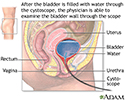
The procedure will take about 5 to 20 minutes. The urethra is cleansed. A numbing medicine is applied to the skin lining the inside of the urethra. This is done without needles. The scope is then inserted through the urethra into the bladder.
Water or salt water (saline) flows through the tube to fill the bladder. As this occurs, you may be asked to describe the feeling. Your answer will give some information about your condition.
As fluid fills the bladder, it stretches the bladder wall. This lets your doctor see the entire bladder wall. You will feel the need to urinate when the bladder is full. However, the bladder must stay full until the exam is finished.
If any tissue looks abnormal, a small sample can be taken (biopsy) through the tube. This sample will be sent to a lab to be tested.
Biopsy
A biopsy is the removal of a small piece of tissue for lab examination.

How to Prepare for the Test
Ask your doctor if you should stop taking any medicines that could thin your blood.
The procedure may be done under anesthesia in a hospital or surgery center. In that case, you will need to have someone take you home afterward.
How the Test will Feel
You may feel slight discomfort when the tube is passed through the urethra into the bladder. You will feel an uncomfortable, strong need to urinate when your bladder is full.
You may feel a quick pinch if a biopsy is taken. After the tube is removed, the urethra may be sore. You may have blood in the urine and a burning sensation during urination for a day or two.
Why the Test is Performed
The test is done to:
- Check for cancer of the bladder or urethra
Cancer
Cancer is the uncontrolled growth of abnormal cells in the body. Cancerous cells are also called malignant cells.
 ImageRead Article Now Book Mark Article
 - Diagnose the cause of blood in the urine
- Diagnose the cause of problems passing urine
- Diagnose the cause of repeated bladder infections
- Help determine the cause of pain during urination
Pain during urination
Painful urination is any pain, discomfort, or burning sensation when passing urine.
 ImageRead Article Now Book Mark Article
Normal Results
The bladder wall should look smooth. The bladder should be of normal size, shape, and position. There should be no blockages, growths, or stones.
What Abnormal Results Mean
The abnormal results could indicate:
-
Bladder cancer
Bladder cancer
Bladder cancer is a cancer that starts in the bladder. The bladder is the body part that holds and releases urine. It is in the center of the lower...
 ImageRead Article Now Book Mark Article
 -
Bladder stones (calculi)
Bladder stones
Bladder stones are hard buildups of minerals. These form in the urinary bladder.
 ImageRead Article Now Book Mark Article
 - Bladder wall decompression
- Chronic urethritis or cystitis
Urethritis
Urethritis is inflammation (swelling and irritation) of the urethra. The urethra is the tube that carries urine from the body.
 ImageRead Article Now Book Mark Article
 - Scarring of the urethra (called a stricture)
- Congenital (present at birth) abnormality
-
Cysts
Cysts
A cyst is a closed pocket or pouch of tissue. It can be filled with air, fluid, pus, or other material.
 ImageRead Article Now Book Mark Article
 -
Diverticula of the bladder or urethra
Diverticula
Diverticula are small, bulging sacs or pouches that form on the inner wall of the intestine. Diverticulitis occurs when these pouches become inflame...
 ImageRead Article Now Book Mark Article
 - Foreign material in the bladder or urethra
Some other possible diagnoses may be:
-
Irritable bladder
Irritable bladder
Urge incontinence occurs when you have a strong, sudden need to urinate that is difficult to delay. The bladder then squeezes, or spasms, and you ma...
 ImageRead Article Now Book Mark Article
 - Polyps
- Prostate problems, such as bleeding, enlargement, or blockage
Enlargement
The prostate is a gland that produces some of the fluid that carries sperm during ejaculation. The prostate gland surrounds the urethra, the tube th...
 ImageRead Article Now Book Mark Article
 -
Traumatic injury of the bladder and urethra
Traumatic injury of the bladder and ure...
Traumatic injury of the bladder and urethra involves damage caused by an outside force.
 ImageRead Article Now Book Mark Article
 -
Ulcer
Ulcer
An ulcer is a crater-like sore on the skin or mucous membrane. Ulcers form when the top layers of skin or tissue have been removed. They can occur ...
 ImageRead Article Now Book Mark Article
 -
Urethral strictures
Urethral strictures
Urethral stricture is an abnormal narrowing of the urethra. The urethra is the tube that carries urine out of the body from the bladder.
 ImageRead Article Now Book Mark Article
Risks
There is a slight risk for excess bleeding when a biopsy is taken.
Other risks include:
-
Bladder infection
Bladder infection
A urinary tract infection, or UTI, is an infection of the urinary tract. The infection can occur at different points in the urinary tract, including...
 ImageRead Article Now Book Mark Article
 - Rupture of the bladder wall
Considerations
Drink 4 to 6 glasses of water per day after the procedure.
You may notice a small amount of blood in your urine after this procedure. If the bleeding continues after you urinate 3 times, contact your doctor.
Contact your doctor if you develop any of these signs of infection:
- Chills
- Fever
- Pain
- Reduced urine output
References
Duty BD, Conlin MJ. Principles of urologic endoscopy. In: Partin AW, Dmochowski RR, Kavoussi LR, Peters CA, eds. Campbell-Walsh-Wein Urology. 12th ed. Philadelphia, PA: Elsevier; 2021:chap 13.
National Institute of Diabetes and Digestive and Kidney Diseases website. Cystoscopy & ureteroscopy. www.niddk.nih.gov/health-information/diagnostic-tests/cystoscopy-ureteroscopy. Updated July 2021. Accessed June 27, 2024.
Taylor JM, Smith TG, Coburn M. Urologic surgery. In: Townsend CM Jr, Beauchamp RD, Evers BM, Mattox KL, eds. Sabiston Textbook of Surgery. 21st ed. Philadelphia, PA: Elsevier; 2022:chap 74.
-
Cystoscopy - illustration
Cystoscopy is a procedure that uses a flexible fiber optic scope inserted through the urethra into the urinary bladder. The physician fills the bladder with water and inspects the interior of the bladder. The image seen through the cystoscope may also be viewed on a color monitor and recorded on videotape for later evaluation.
Cystoscopy
illustration
Review Date: 5/17/2024
Reviewed By: Sovrin M. Shah, MD, Associate Professor, Department of Urology, The Icahn School of Medicine at Mount Sinai, New York, NY. Review provided by VeriMed Healthcare Network. Also reviewed by David C. Dugdale, MD, Medical Director, Brenda Conaway, Editorial Director, and the A.D.A.M. Editorial team.


