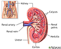
Kidney biopsy
Renal biopsy; Biopsy - kidneyA kidney biopsy is the removal of a small piece of kidney tissue for examination.
How the Test is Performed
A kidney biopsy is done in the hospital. The two most common ways to do a kidney biopsy are percutaneous and open. These are described below.
Percutaneous biopsy. Percutaneous means through the skin. Most kidney biopsies are done this way. The procedure is usually done in the following way:
- You may receive medicine to make you drowsy.
- You lie on your stomach. If you have a transplanted kidney, you lie on your back.
- The health care provider marks the spot on the skin where the biopsy needle is inserted.
- The skin is cleaned.
- Numbing medicine (anesthetic) is injected under the skin near the kidney area.
- The provider makes a tiny cut in the skin. Ultrasound images are used to find the proper location. Sometimes another imaging method, such as CT, is used.
Ultrasound
Ultrasound uses high-frequency sound waves to make images of organs and structures inside the body.
 ImageRead Article Now Book Mark Article
CT
A computed tomography (CT) scan is an imaging method that uses x-rays to create pictures of cross-sections of the body. Related tests include:Abdomin...
 ImageRead Article Now Book Mark Article
 - The provider inserts a biopsy needle through the skin to the surface of the kidney. You are asked to take and hold a deep breath as the needle goes into the kidney.
- If the provider is not using ultrasound guidance, you may be asked to take several deep breaths. This allows the provider to know the needle is in place.
- The needle may be inserted more than once if more than one tissue sample is needed.
- The needle is removed. Pressure is applied to the biopsy site to stop any bleeding.
- After the procedure, a bandage is applied to the biopsy site.
Open biopsy. In some cases, your provider may recommend a surgical (open) biopsy. This method is used when a larger piece of tissue is needed or a percutaneous needle biopsy cannot be done safely.
- You receive medicine (anesthesia) that allows you to sleep and be pain-free.
- The surgeon makes a small surgical cut (incision).
- The surgeon locates the part of the kidney from which the biopsy tissue needs to be taken. The tissue is removed.
- The incision is closed with stitches (sutures).
After percutaneous or open biopsy, you will likely stay in the hospital for at least 12 hours. You will receive pain medicines and fluids by mouth or through a vein (IV). Your urine will be checked for heavy bleeding. A small amount of bleeding is normal after a biopsy.
Follow instructions about caring for yourself after the biopsy. This may include not lifting anything heavier than 10 pounds (4.5 kilograms) for 2 weeks after the biopsy.
How to Prepare for the Test
Tell your provider:
- About medicines you are taking, including vitamins and supplements, herbal remedies, and over-the-counter medicines
- If you have any allergies
- If you have bleeding problems or if you take blood-thinning medicines such as warfarin (Coumadin), clopidogrel (Plavix), dipyridamole (Persantine), fondaparinux (Arixtra), apixaban (Eliquis), dabigatran (Pradaxa), or aspirin
- If you are or think you might be pregnant
How the Test will Feel
Numbing medicine is used, so the pain during the procedure is often slight. The numbing medicine may burn or sting when first injected.
After the procedure, the area may feel tender or sore for a few days.
You may see bright, red blood in your urine during the first 24 hours after the test. If the bleeding lasts longer, tell your provider.
Why the Test is Performed
Your provider may order a kidney biopsy if you have:
- An unexplained drop in kidney function
-
Blood in the urine that does not go away
Blood in the urine
Blood in your urine is called hematuria. The amount may be very small and only detected with urine tests or under a microscope. In other cases, the...
 ImageRead Article Now Book Mark Article
 -
Protein in the urine found during a urine test
Protein in the urine
The urine protein dipstick test measures the presence of all proteins, including albumin, in a urine sample. Albumin and protein can each also be mea...
 ImageRead Article Now Book Mark Article
 - A transplanted kidney, which needs to be monitored using a biopsy
Normal Results
A normal result is when the kidney tissue shows normal structure.
What Abnormal Results Mean
An abnormal result means there are changes in the kidney tissue. This may be due to:
- Infection
- Poor blood flow through the kidney
- Connective tissue diseases such as systemic lupus erythematosus
Systemic lupus erythematosus
Systemic lupus erythematosus (SLE) is an autoimmune disease. In this disease, the immune system of the body mistakenly attacks healthy tissue. It c...
 ImageRead Article Now Book Mark Article
 - Other diseases that may be affecting the kidney, such as diabetes
- Kidney transplant rejection, if you had a transplant
Transplant rejection
Transplant rejection is a process in which a transplant recipient's immune system attacks the transplanted organ or tissue.
 ImageRead Article Now Book Mark Article
Risks
Risks include:
- Bleeding from the kidney (in rare cases, may require a blood transfusion)
- Bleeding into the muscle, which might cause soreness
- Infection (small risk)
References
Salama AD, Cook HT. The renal biopsy. In: Yu ASL, Chertow GM, Luyckx VA, Marsden PA, Karl S, Philip AM, Taal MW, eds. Brenner and Rector's The Kidney. 11th ed. Philadelphia, PA: Elsevier; 2020:chap 26.
Topham PS, MacGinley. Kidney biopsy. In: Johnson RJ, Floege J, Tonelli M, eds. Comprehensive Clinical Nephrology. 7th ed. Philadelphia, PA: Elsevier; 2024:chap 7.
-
Kidney anatomy - illustration
The kidneys are responsible for removing wastes from the body, regulating electrolyte balance and blood pressure, and the stimulation of red blood cell production.
Kidney anatomy
illustration
-
Kidney - blood and urine flow - illustration
This is the typical appearance of the blood vessels (vasculature) and urine flow pattern in the kidney. The blood vessels are shown in red and the urine flow pattern in yellow.
Kidney - blood and urine flow
illustration
-
Renal biopsy - illustration
In renal biopsy, a small sample of kidney tissue is removed with a needle. The test is sometimes used to evaluate a transplanted kidney. It is also used to evaluate an unexplained decrease in kidney function, persistent blood in the urine, or protein in the urine.
Renal biopsy
illustration
-
Kidney anatomy - illustration
The kidneys are responsible for removing wastes from the body, regulating electrolyte balance and blood pressure, and the stimulation of red blood cell production.
Kidney anatomy
illustration
-
Kidney - blood and urine flow - illustration
This is the typical appearance of the blood vessels (vasculature) and urine flow pattern in the kidney. The blood vessels are shown in red and the urine flow pattern in yellow.
Kidney - blood and urine flow
illustration
-
Renal biopsy - illustration
In renal biopsy, a small sample of kidney tissue is removed with a needle. The test is sometimes used to evaluate a transplanted kidney. It is also used to evaluate an unexplained decrease in kidney function, persistent blood in the urine, or protein in the urine.
Renal biopsy
illustration
Review Date: 8/28/2023
Reviewed By: Walead Latif, MD, Nephrologist and Clinical Associate Professor, Rutgers Medical School, Newark, NJ. Review provided by VeriMed Healthcare Network. Also reviewed by David C. Dugdale, MD, Medical Director, Brenda Conaway, Editorial Director, and the A.D.A.M. Editorial team.


