Endometrial biopsy
Biopsy - endometriumEndometrial biopsy is the removal of a small piece of tissue from the lining of the uterus (endometrium) for examination.
Biopsy
A biopsy is the removal of a small piece of tissue for lab examination.

How the Test is Performed
This procedure may be done with or without anesthesia. This is medicine that allows you to sleep during the procedure.
- You lie on your back with your feet in stirrups, similar to having a pelvic exam.
- Your health care provider gently inserts an instrument (speculum) into the vagina to hold it open so that your cervix can be viewed. The cervix is cleaned with a special liquid. Numbing medicine may be applied to the cervix.
Cervix
The cervix is the lower end of the womb (uterus). It is at the top of the vagina. It is about 2. 5 to 3. 5 centimeters (1 to 1. 3 inches) long. Th...
 ImageRead Article Now Book Mark Article
 - The cervix may then be gently grasped with an instrument to hold the uterus steady. Another instrument may be needed to gently stretch the cervical opening if there is tightness.
- An instrument is gently passed through the cervix into the uterus to collect the tissue sample.
- The tissue sample and instruments are removed.
- The tissue is sent to a lab. There, it is examined under a microscope.
- If you had anesthesia for the procedure, you are taken to a recovery area. Nurses will make sure you are comfortable. After you wake up and have no problems from the anesthesia and procedure, you are allowed to go home.
How to Prepare for the Test
Before the test:
- Tell your provider about all the medicines you take. These include blood thinners such as warfarin, clopidogrel, and aspirin.
- You may be asked to have a test to make sure you are not pregnant.
- In the 2 days before the procedure, do not use creams or other medicines in the vagina.
- Do NOT douche. (You should never douche. Douching can cause infection of the vagina or uterus.)
- Ask your provider if you should take pain medicine, such as ibuprofen or acetaminophen, just before the procedure.
How the Test will Feel
The instruments may feel cold. You may feel some cramping when the cervix is grasped. You may have some mild cramping as the instruments enter the uterus and the sample is collected. The discomfort is mild, though for some women it can be severe. However, the duration of the test and the pain are short.
Why the Test is Performed
The test is done to find the cause of:
- Abnormal menstrual periods (heavy, prolonged, or irregular bleeding)
- Bleeding after menopause
Menopause
Menopause is the time in a woman's life when her periods (menstruation) stop. Most often, it is a natural, normal body change that occurs between ag...
 ImageRead Article Now Book Mark Article
 - Bleeding from taking hormone therapy medicines
- Thickened uterine lining seen on ultrasound
-
Endometrial cancer
Endometrial cancer
Endometrial cancer is cancer that starts in the endometrium, the lining of the uterus (womb).
 ImageRead Article Now Book Mark Article
Normal Results
The biopsy is normal if the cells in the sample are not abnormal.
What Abnormal Results Mean
Abnormal menstrual periods may be caused by:
-
Uterine fibroids
Uterine fibroids
Uterine fibroids are tumors that grow in a woman's womb (uterus). These growths are typically not cancerous (benign), and do not become cancerous....
 ImageRead Article Now Book Mark Article
 - Fingerlike growths in the uterus (uterine polyps)
- Infection
- Hormone imbalance
- Endometrial cancer or precancer (hyperplasia)
Hyperplasia
Hyperplasia is increased cell production in a normal tissue or organ. Hyperplasia may be a sign of abnormal or precancerous changes. This is called...
 ImageRead Article Now Book Mark Article
Other conditions under which the test may be performed:
- Abnormal bleeding if a woman is taking the breast cancer medicine tamoxifen
- Abnormal bleeding due to changes in hormone levels (anovulatory bleeding)
Anovulatory bleeding
Abnormal uterine bleeding (AUB) is bleeding from the uterus that is longer than usual or that occurs at an irregular time. Bleeding may be heavier o...
 ImageRead Article Now Book Mark Article
Risks
Risks for endometrial biopsy include:
- Infection
- Causing a hole in (perforating) the uterus or tearing the cervix (rarely occurs)
- Prolonged bleeding
- Slight spotting and mild cramping for a few days
References
Beard JM, Osborn J. Common office procedures. In: Rakel RE, Rakel DP, eds. Textbook of Family Medicine. 9th ed. Philadelphia, PA: Elsevier Saunders; 2016:chap 28.
Soliman PT, Lu KH. Malignant diseases of the uterus: endometrial hyperplasia, endometrial carcinoma, sarcoma: diagnosis and management. In: Gershenson DM, Lentz GM, Valea FA, Lobo RA, eds. Comprehensive Gynecology. 8th ed. Philadelphia, PA: Elsevier; 2022:chap 32.
-
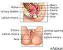
Female reproductive anatomy - illustration
Internal structures of the female reproductive anatomy include the uterus, ovaries, and cervix. External structures include the labium minora and majora, the vagina and the clitoris.
Female reproductive anatomy
illustration
-
Endometrial biopsy - illustration
The mucosal lining of the cavity of the uterus is called the endometrium. It is this lining which undergoes changes over the course of the monthly menstrual cycle, sloughes off and becomes part of the menses. A biopsy of the endometrium is used to check for disease or problems of fertility.
Endometrial biopsy
illustration
-
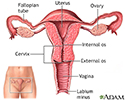
Uterus - illustration
The uterus is a hollow muscular organ located in the female pelvis between the bladder and rectum. The ovaries produce the eggs that travel through the fallopian tubes. Once the egg has left the ovary it can be fertilized and implant itself in the lining of the uterus. The main function of the uterus is to nourish the developing fetus prior to birth.
Uterus
illustration
-
Endometrial biopsy - illustration
An endometrial biopsy is a procedure in which a tissue sample is obtained from the endometrium (the inside lining of the uterus) and is then observed under a microscope. The tissue is thoroughly examined for any cell abnormalities or cancer. The test also helps determine the cause of abnormal menstrual periods, and can be used to screen for endometrial cancer. The test is sometimes used as part of the diagnostic work-up of women who have been unable to become pregnant.
Endometrial biopsy
illustration
-
Female reproductive anatomy - illustration
Internal structures of the female reproductive anatomy include the uterus, ovaries, and cervix. External structures include the labium minora and majora, the vagina and the clitoris.
Female reproductive anatomy
illustration
-
Endometrial biopsy - illustration
The mucosal lining of the cavity of the uterus is called the endometrium. It is this lining which undergoes changes over the course of the monthly menstrual cycle, sloughes off and becomes part of the menses. A biopsy of the endometrium is used to check for disease or problems of fertility.
Endometrial biopsy
illustration
-
Uterus - illustration
The uterus is a hollow muscular organ located in the female pelvis between the bladder and rectum. The ovaries produce the eggs that travel through the fallopian tubes. Once the egg has left the ovary it can be fertilized and implant itself in the lining of the uterus. The main function of the uterus is to nourish the developing fetus prior to birth.
Uterus
illustration
-
Endometrial biopsy - illustration
An endometrial biopsy is a procedure in which a tissue sample is obtained from the endometrium (the inside lining of the uterus) and is then observed under a microscope. The tissue is thoroughly examined for any cell abnormalities or cancer. The test also helps determine the cause of abnormal menstrual periods, and can be used to screen for endometrial cancer. The test is sometimes used as part of the diagnostic work-up of women who have been unable to become pregnant.
Endometrial biopsy
illustration
Review Date: 8/23/2023
Reviewed By: LaQuita Martinez, MD, Department of Obstetrics and Gynecology, Emory Johns Creek Hospital, Alpharetta, GA. Also reviewed by David C. Dugdale, MD, Medical Director, Brenda Conaway, Editorial Director, and the A.D.A.M. Editorial team.


