
Bone lesion biopsy
Bone biopsy; Biopsy - boneA bone lesion biopsy is the removal of a piece of bone or bone marrow for examination.
How the Test is Performed
The test is done in the following way:
- An x-ray, CT or MRI scan is likely used to guide the exact placement of the biopsy instrument.
x-ray
X-rays are a type of electromagnetic radiation, just like visible light. An x-ray machine sends individual x-ray waves through the body. The images...
 ImageRead Article Now Book Mark Article
CT
A computed tomography (CT) scan is an imaging method that uses x-rays to create pictures of cross-sections of the body. Related tests include:Abdomin...
 ImageRead Article Now Book Mark Article
MRI
A magnetic resonance imaging (MRI) scan is an imaging test that uses powerful magnets and radio waves to create pictures of the body. It does not us...
 ImageRead Article Now Book Mark Article
 - Your surgeon applies a numbing medicine (local anesthetic) to the area.
- A small cut is then made in the skin.
- A special drill needle is often used. This needle is gently inserted through the cut, then pushed and twisted into the bone.
- Once the sample is obtained, the needle is twisted out.
- Pressure is applied to the site. Once bleeding stops, stitches are applied, and covered with a bandage.
- The sample is sent to a lab for examination.
Bone biopsy may also be done under general anesthesia to remove a larger sample. Then surgery to remove the bone can be done if the biopsy exam shows that there is an abnormal growth or cancer.
Cancer
Cancer is the uncontrolled growth of abnormal cells in the body. Cancerous cells are also called malignant cells.

How to Prepare for the Test
Follow your surgeon's instructions on how to prepare. This may include not eating and drinking for several hours before the procedure. If you take any blood thinners, please make sure you stop them ahead of the procedure.
How the Test will Feel
With a needle biopsy, you may feel some discomfort and pressure, even though a local anesthetic is used. You must remain still during the procedure.
After the biopsy, the area may be sore or tender for several days.
Why the Test is Performed
The most common reasons for bone lesion biopsy are to tell the difference between cancerous and noncancerous bone tumors and to identify other bone or bone marrow problems. It may be performed on people with bone pain and tenderness, particularly if x-ray, CT scan, or other testing reveals a problem.
Bone pain
Bone pain or tenderness is aching or other discomfort in one or more bones.

Normal Results
No abnormal bone tissue is found.
What Abnormal Results Mean
An abnormal result may be any of the following problems.
Benign (noncancerous) bone tumors, such as:
Benign
Benign refers to a condition, tumor, or growth that is not cancerous. This means that it does not spread to other parts of the body. It does not in...

- Bone cyst
Cyst
A cyst is a closed pocket or pouch of tissue. It can be filled with air, fluid, pus, or other material.
 ImageRead Article Now Book Mark Article
 - Fibroma
-
Osteoblastoma
Osteoblastoma
A bone tumor is an abnormal growth of cells within a bone. A bone tumor may be cancerous (malignant) or noncancerous (benign).
ImageRead Article Now Book Mark Article -
Osteoid osteoma
Osteoid osteoma
A bone tumor is an abnormal growth of cells within a bone. A bone tumor may be cancerous (malignant) or noncancerous (benign).
ImageRead Article Now Book Mark Article
Cancerous tumors, such as:
-
Ewing sarcoma
Ewing sarcoma
Ewing sarcoma is a malignant bone tumor that forms in the bone or soft tissue around it. It affects mostly teens and young adults.
ImageRead Article Now Book Mark Article -
Multiple myeloma
Multiple myeloma
Multiple myeloma is a blood cancer that starts from a type of white blood cell in the bone marrow called plasma cells. Bone marrow is the soft, spon...
 ImageRead Article Now Book Mark Article
 -
Osteosarcoma
Osteosarcoma
Osteosarcoma is a rare type of cancerous bone tumor that usually develops in teenagers and young adults. It often occurs when a teen is growing rapi...
ImageRead Article Now Book Mark Article - Other types of cancer that may have spread to the bone
Abnormal results may also be due to:
-
Osteitis fibrosa (weak and deformed bone)
Osteitis fibrosa
Osteitis fibrosa is a complication of hyperparathyroidism, a condition in which overactive parathyroid glands cause certain bones to become abnormall...
 ImageRead Article Now Book Mark Article
 -
Osteomalacia (softening of the bones)
Osteomalacia
Osteomalacia is softening of the bones. It most often occurs because of a problem that leads to vitamin D deficiency, which helps your body absorb c...
 ImageRead Article Now Book Mark Article
 -
Osteomyelitis (bone infection)
Osteomyelitis
Osteomyelitis is a bone infection. It is caused by bacteria or other germs.
ImageRead Article Now Book Mark Article - Bone marrow disorders (leukemia or lymphoma)
Leukemia
Leukemia is a type of blood cancer that begins in the bone marrow. Bone marrow is the soft tissue in the center of the bones, where blood cells are ...
 ImageRead Article Now Book Mark Article
Risks
Risks of this procedure may include:
-
Bone fracture
Bone fracture
If more pressure is put on a bone than it can stand, it will split or break. A break of any size is called a fracture. If the broken bone punctures...
ImageRead Article Now Book Mark Article - Bone infection (osteomyelitis)
Osteomyelitis
Osteomyelitis is a bone infection. It is caused by bacteria or other germs.
ImageRead Article Now Book Mark Article - Damage to surrounding tissue
- Discomfort
- Excessive bleeding
- Infection near the biopsy area
A serious risk of this procedure is bone infection. Signs of a bone infection include:
- Fever
- Chills
- Worsening pain
- Redness and swelling of around the biopsy site
- Drainage of pus from the biopsy site
- Difficulty with walking or using your limb
If you have any of these signs, contact your health care provider or surgeon right away.
People with bone disorders who also have blood clotting disorders may have an increased risk of bleeding.
References
Jayaram PR, Yan YY, Mallinson PI, Ouellette HA, Munk PL. Interventional radiologic techniques in the management of bone tumors. In: Heymann D, ed. Bone Cancer. 3rd ed. Philadelphia, PA: Elsevier; 2022:chap 55.
Katsanos K, Sabharwal T, Cazzato RL, Gangi A. Skeletal interventions. In: Adam A, Dixon AK, Gillard JH, Schaefer-Prokop CM, eds. Grainger & Allison's Diagnostic Radiology. 7th ed. Philadelphia, PA: Elsevier; 2021:chap 87.
Schwartz HS, Holt GE, Halpern JL. Bone tumors. In: Townsend CM Jr, Beauchamp RD, Evers BM, Mattox KL, eds. Sabiston Textbook of Surgery. 21st ed. St. Louis, MO: Elsevier; 2022:chap 33.
-
Bone biopsy - illustration
A bone biopsy is performed by making a small incision into the skin. A biopsy needle retrieves a sample of bone and it is sent for examination. The most common reasons for bone lesion biopsy are to distinguish between benign and malignant bone tumors, and to identify other bone abnormalities. Bone biopsy may also be performed to determine the cause of bone pain and tenderness.
Bone biopsy
illustration
-
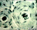
Bone Tissue - illustration
A photomicrograph of bone tissue. Bone tissue is obtained from a bone biopsy and examined under a microscope. This is a picture of how normal tissue appears when magnified.
Bone Tissue
illustration

-
Bone biopsy - illustration
A bone biopsy is performed by making a small incision into the skin. A biopsy needle retrieves a sample of bone and it is sent for examination. The most common reasons for bone lesion biopsy are to distinguish between benign and malignant bone tumors, and to identify other bone abnormalities. Bone biopsy may also be performed to determine the cause of bone pain and tenderness.
Bone biopsy
illustration
-
Bone Tissue - illustration
A photomicrograph of bone tissue. Bone tissue is obtained from a bone biopsy and examined under a microscope. This is a picture of how normal tissue appears when magnified.
Bone Tissue
illustration
Review Date: 8/27/2024
Reviewed By: C. Benjamin Ma, MD, Professor, Chief, Sports Medicine and Shoulder Service, UCSF Department of Orthopaedic Surgery, San Francisco, CA. Also reviewed by David C. Dugdale, MD, Medical Director, Brenda Conaway, Editorial Director, and the A.D.A.M. Editorial team.
 All rights reserved.