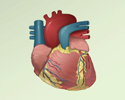
Heart CT scan
CAT scan - heart; Computed axial tomography scan - heart; Computed tomography scan - heart; Calcium scoring; Multi-detector CT scan - heart; Electron beam computed tomography - heart; Agatston score; Coronary calcium scanA computed tomography (CT) scan of the heart is an imaging method that uses x-rays to create detailed pictures of the heart and its blood vessels.
- This test is called a coronary calcium scan when it is done to see if you have a buildup of calcium in your heart arteries (also called coronary arteries).
- It is called CT angiography if it is done to look at the arteries that bring blood to your heart. This test evaluates if there is narrowing or a blockage in those arteries.
- The test is sometimes done in combination with scans of the aorta or pulmonary arteries to look for problems with those structures.
Aorta
Aortic angiography is a procedure that uses a special dye and x-rays to see how blood flows through the aorta. The aorta is the large artery that ca...
 ImageRead Article Now Book Mark Article
Pulmonary arteries
Pulmonary angiography is a test to see how blood flows through the lung. Angiography is an imaging test that uses x-rays and a special dye to see th...
 ImageRead Article Now Book Mark Article
How the Test is Performed
You will be asked to lie on a narrow table that slides into the center of the CT scanner.
- You will lie on your back with your head and feet outside the scanner on either end.
- Small patches, called electrodes are put on your chest and connected to a machine that records your heart's electrical activity. You may be given medicine to slow your heart rate.
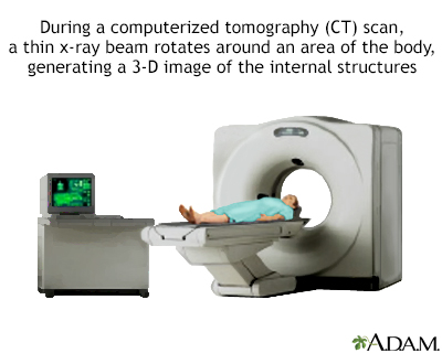
- Once you are inside the scanner, the machine's x-ray beam rotates around you.
A computer creates separate images of the body area, called slices.
- These images can be stored, viewed on a monitor, or printed on film.
- 3D (three-dimensional) models of the heart can be created.
You must be still during the exam because movement causes blurred images. You may be told to hold your breath for short periods of time.
The entire scan should only take about 10 minutes.
How to Prepare for the Test
Certain exams require a special dye, called contrast, to be delivered into the body before the test starts. Contrast helps certain areas show up better on the x-rays.
- Contrast can be given through a vein (IV) in your hand or forearm. If contrast is used, you may also be asked not to eat or drink anything for 4 to 6 hours before the test.
Before receiving the contrast:
- Let your health care provider know if you have ever had a reaction to contrast or any medicines. You may need to take medicines before the test in order to safely receive this substance.
- Tell your provider about all your medicines, because you may be asked to hold some, such as the diabetes medicine metformin (Glucophage and others), prior to the test.
- Let your provider know if you have kidney problems. The contrast material can cause kidney function to worsen.
If you weigh more than 300 pounds (136 kilograms), find out if the CT machine has a weight limit. Too much weight can cause damage to the scanner's working parts.
You will be asked to remove jewelry and wear a hospital gown during the study.
How the Test will Feel
Some people may have discomfort from lying on the hard table.
Contrast given through an IV may cause a:
- Slight burning sensation
- Metallic taste in the mouth
- Warm flushing of the body
These sensations are normal and usually go away within a few seconds.
Why the Test is Performed
CT rapidly creates detailed pictures of the heart and its arteries. The test may diagnose or detect:
- Plaque buildup in the coronary arteries to determine your risk for heart disease
-
Congenital heart disease (heart problems that are present at birth)
Congenital heart disease
Congenital heart disease (CHD) is a problem with the heart's structure and function that is present at birth.
 ImageRead Article Now Book Mark Article
 - Problems with the heart valves
- Blockage of the arteries that supply the heart
- Tumors or masses of the heart
- Pumping function of the heart
Normal Results
Results are considered normal if the heart and arteries being examined are normal in appearance.
Your "calcium score" is based on the amount of calcium (plaque) found in the arteries of your heart.
- The test is normal (negative) if your calcium score is 0. This means the chance of having a heart attack over the next several years is very low.
- If the calcium score is very low, you are unlikely to have coronary artery disease.
Coronary artery disease
Coronary heart disease is a narrowing of the blood vessels that supply blood and oxygen to the heart. Coronary heart disease (CHD) is also called co...
 ImageRead Article Now Book Mark Article
What Abnormal Results Mean
Abnormal results may be due to:
-
Aneurysm
Aneurysm
An aneurysm is an abnormal widening or ballooning of a part of an artery due to weakness in the wall of the blood vessel.
 ImageRead Article Now Book Mark Article
 - Congenital heart disease
- Coronary artery disease
- Heart valve problems
- Inflammation of the covering around the heart (pericarditis)
Pericarditis
Pericarditis is a condition in which the sac-like covering around the heart (pericardium) becomes inflamed.
 ImageRead Article Now Book Mark Article
 - Narrowing of one or more coronary arteries (coronary artery stenosis)
- Tumors or other masses of the heart or surrounding areas
Calcium scores above 0 indicate the presence of plaque in your arteries:
- 1 to 9 - minimal
- 10 to 99 - mild
- 100 to 299 - moderate
- 300 to 999 - severe
- 1000 or above - extreme
If your calcium score is high:
- It means you have calcium buildup in the walls of your coronary arteries. This is usually a sign of atherosclerosis, or hardening of the arteries.
Atherosclerosis
Atherosclerosis, sometimes called "hardening of the arteries," occurs when fat, cholesterol, and other substances build up in the walls of arteries. ...
 ImageRead Article Now Book Mark Article
 - The higher your score, the more severe this problem may be.
- Talk to your provider about lifestyle or other changes you can make to decrease the risk for heart disease.
Risks
Risks for CT scans include:
- Being exposed to radiation
- Allergic reaction to contrast dye
CT scans do expose you to more radiation than regular x-rays. Having many x-rays or CT scans over time may increase your risk for cancer. However, the risk from any one scan is small. You and your provider should weigh this risk against the benefits of getting a correct diagnosis for a medical problem.
Some people have allergies to contrast dye. Let your provider know if you have ever had an allergic reaction to injected contrast dye.
- The most common type of contrast given into a vein contains iodine. If a person with an iodine allergy is given this type of contrast, nausea or vomiting, sneezing, itching, or hives may occur.
Nausea or vomiting
Nausea is feeling an urge to vomit. It is often called "being sick to your stomach. "Vomiting or throwing-up forces the contents of the stomach up t...
 ImageRead Article Now Book Mark Article
Sneezing
A sneeze is a sudden, forceful, uncontrolled burst of air through the nose and mouth.
 ImageRead Article Now Book Mark Article
Itching
Itching is a tingling or irritation of the skin that makes you want to scratch the area. Itching may occur all over the body or only in one location...
 ImageRead Article Now Book Mark Article
Hives
Hives are raised, usually itchy, red bumps (welts) on the surface of the skin. They can be an allergic reaction to food or medicine. They can also ...
 ImageRead Article Now Book Mark Article
 - If you absolutely must be given such contrast, you may need to take steroids (such as prednisone) or antihistamines (such as diphenhydramine) before the test. You may also need to take a histamine blocker (such as ranitidine).
- The kidneys help remove iodine out of the body. Those with kidney disease or diabetes may need to receive extra fluids after the test to help flush the iodine out of the body.
Rarely, the dye may cause a life-threatening allergic response called anaphylaxis. If you have any trouble breathing during the test, you should notify the scanner operator immediately. Scanners come with an intercom and speakers, so the operator can hear you at all times.
Anaphylaxis
Anaphylaxis is a life-threatening type of allergic reaction.

References
Arnett DK, Blumenthal RS, Albert MA, et al. 2019 ACC/AHA Guideline on the Primary Prevention of Cardiovascular Disease: A Report of the American College of Cardiology/American Heart Association Task Force on Clinical Practice Guidelines. Circulation. 2019;140(11):e596-e646. PMID: 30879355 pubmed.ncbi.nlm.nih.gov/30879355/.
Blankstein R. Cardiac computed tomography. In: Libby P, Bonow RO, Mann DL, Tomaselli GF, Bhatt DL, Solomon SD, eds. Braunwald's Heart Disease: A Textbook of Cardiovascular Medicine. 12th ed. Philadelphia, PA: Elsevier; 2022:chap 20.
Doherty JU, Kort S, Mehran R, et al. ACC/AATS/AHA/ASE/ASNC/HRS/SCAI/SCCT/SCMR/STS 2019 appropriate use criteria for multimodality imaging in the assessment of cardiac structure and function in nonvalvular heart disease: a report of the American College of Cardiology Appropriate Use Criteria Task Force, American Association for Thoracic Surgery, American Heart Association, American Society of Echocardiography, American Society of Nuclear Cardiology, Heart Rhythm Society, Society for Cardiovascular Angiography and Interventions, Society of Cardiovascular Computed Tomography, Society for Cardiovascular Magnetic Resonance, and Society of Thoracic Surgeons. .J Am Coll Cardiol. 2019;73(4):488-516. PMID: 30630640 pubmed.ncbi.nlm.nih.gov/30630640/.
Kramer CM, Dilsizian V, Hagspiel KD. Noninvasive cardiac imaging. In: Goldman L, Cooney KA, eds. Goldman-Cecil Medicine. 27th ed. Philadelphia, PA: Elsevier; 2024:chap 44.
-
CT scan - illustration
CT stands for computerized tomography. In this procedure, a thin X-ray beam is rotated around the area of the body to be visualized. Using very complicated mathematical processes called algorithms, the computer is able to generate a 3-D image of a section through the body. CT scans are very detailed and provide excellent information for the physician.
CT scan
illustration
Review Date: 5/5/2025
Reviewed By: Michael A. Chen, MD, PhD, Associate Professor of Medicine, Division of Cardiology, Harborview Medical Center, University of Washington Medical School, Seattle, WA. Also reviewed by David C. Dugdale, MD, Medical Director, Brenda Conaway, Editorial Director, and the A.D.A.M. Editorial team.


